Claessens P, Claessens C, Claessens J, Claessens M Strain Imaging: Key to the Specific Left Ventricular Diastolic Properties in Endurance Trained Athletes Journal of Clinical and Basic Cardiology 2003; 6 (1-4): 35-40 PDF Summary Overview
| ||||||||||||||||
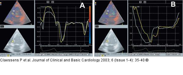
Figure/Graphic 5a-b: Athlet - A-Wave A: Velocity curve (tissue Doppler imaging): localisation of the end of the A-wave and determination the time on the end of the A-wave at the basal and middle septum in the long apical axis; B: Strain curve: measurement of the strain value at the time of the end of the A-wave at the basal and middle septum in the long apical axis. |

Figure/Graphic 5a-b: Athlet - A-Wave
A: Velocity curve (tissue Doppler imaging): localisation of the end of the A-wave and determination the time on the end of the A-wave at the basal and middle septum in the long apical axis; B: Strain curve: measurement of the strain value at the time of the end of the A-wave at the basal and middle septum in the long apical axis. |


