Perneczky G, Loyoddin M, Schappelwein H, Sherif C Spinal Meningiomas: A Comprehensive Overview and Own Experience European Association of NeuroOncology Magazine 2013; 3 (3): 118-121 PDF Summary Overview
| ||||
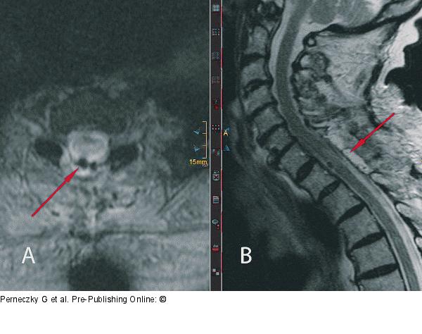
Figure/Graphic 2: Spinal Menigioma Postoperative MR scans: (a) T1-weighted contrast-enhanced MR images: the spinal cord (red arrow) is in normal location. No tumour remnants are visible. (b) T2-weighted native MR scan: the surgical dorsal approach and complete tumour removal are documented. |

Figure/Graphic 2: Spinal Menigioma
Postoperative MR scans: (a) T1-weighted contrast-enhanced MR images: the spinal cord (red arrow) is in normal location. No tumour remnants are visible. (b) T2-weighted native MR scan: the surgical dorsal approach and complete tumour removal are documented. |


