Mundigler G, Zehetgruber M Tissue Doppler Imaging: Myocardial Velocities and Strain - Are there Clinical Applications? Journal of Clinical and Basic Cardiology 2002; 5 (2): 125-132 PDF Summary Overview
| ||||
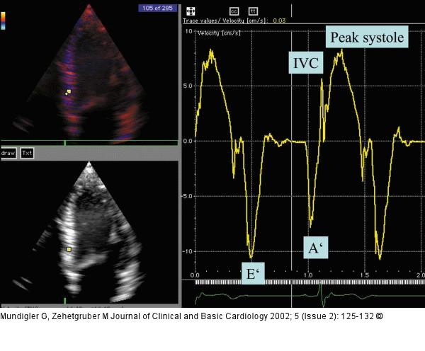
Figure/Graphic 2: Gewebe-Doppler Tissue Doppler velocity curve in a healthy volunteer. Apical 4-chamber view. The sample volume is positioned in the basal inferior septum. The initial positive excursion represents the isovolaemic contraction phase (IVC), followed by the systole. The negative waves represent early (E') and late (A') diastole. |

Figure/Graphic 2: Gewebe-Doppler
Tissue Doppler velocity curve in a healthy volunteer. Apical 4-chamber view. The sample volume is positioned in the basal inferior septum. The initial positive excursion represents the isovolaemic contraction phase (IVC), followed by the systole. The negative waves represent early (E') and late (A') diastole. |


