Mundigler G, Zehetgruber M Tissue Doppler Imaging: Myocardial Velocities and Strain - Are there Clinical Applications? Journal of Clinical and Basic Cardiology 2002; 5 (2): 125-132 PDF Summary Overview
| ||||
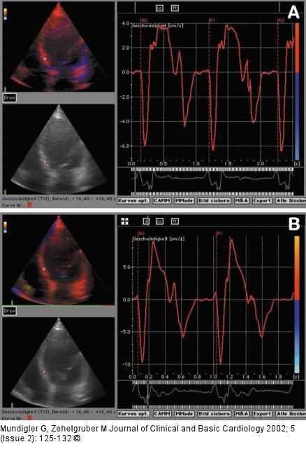
Figure/Graphic 3a-b: Gewebe-Doppler a) TDI-velocity curves in a patient with ischaemic cardiomyopathy. In contrast to Fig. 2 there is reduced velocity (3.8 cm/s) at rest due to hypokinesis in the inferior basal septum. b) During peak dobutamine stress there is a significant increase up to approximately 10 cm/s, indicating contractile reserve. Note the reversal of E'/A' indicating diastolic dysfunction. |

Figure/Graphic 3a-b: Gewebe-Doppler
a) TDI-velocity curves in a patient with ischaemic cardiomyopathy. In contrast to Fig. 2 there is reduced velocity (3.8 cm/s) at rest due to hypokinesis in the inferior basal septum. b) During peak dobutamine stress there is a significant increase up to approximately 10 cm/s, indicating contractile reserve. Note the reversal of E'/A' indicating diastolic dysfunction. |


