Bartel T, Caspari G, Erbel R, Mueller S Intracardiac Echocardiography - Technology and Clinical Role Journal of Clinical and Basic Cardiology 2002; 5 (2): 133-137 PDF Summary Overview
| ||||||||||||||||
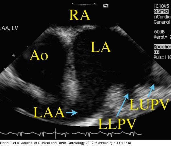
Figure/Graphic 3: Intrakardialer Ultraschall Left atrium, left atrial appendage, and left pulmonary veins demonstrated with the AcuNav(TM)-catheter being placed in the right atrium and lined up with the interatrial septum. Ao = aorta; LAA = left atrial appendage; LA = left atrium; LLPV = left lower pulmonary vein; LUPV = left upper pulmonary vein; RA = right atrium |

Figure/Graphic 3: Intrakardialer Ultraschall
Left atrium, left atrial appendage, and left pulmonary veins demonstrated with the AcuNav(TM)-catheter being placed in the right atrium and lined up with the interatrial septum. Ao = aorta; LAA = left atrial appendage; LA = left atrium; LLPV = left lower pulmonary vein; LUPV = left upper pulmonary vein; RA = right atrium |


