Bartel T, Caspari G, Erbel R, Mueller S Intracardiac Echocardiography - Technology and Clinical Role Journal of Clinical and Basic Cardiology 2002; 5 (2): 133-137 PDF Summary Overview
| ||||||||||||||||||
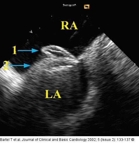
Figure/Graphic 5: Intrakardiale Echokardiographie Device closure of a patent foramen ovale guided by intracardiac echocardiography with the AcuNav )(TM)-catheter placed in the right atrium and retroflexed into a short-axis orientation. Typical attitude before the Amplatzer (TM)-PFO occluder is removed from the cable. LA = left atrium; RA = right atrium; 1, right umbrella of Amplatzer (TM)-PFO occluder; 2, left umbrella of Amplatzer (TM)-PFO occluder |

Figure/Graphic 5: Intrakardiale Echokardiographie
Device closure of a patent foramen ovale guided by intracardiac echocardiography with the AcuNav )(TM)-catheter placed in the right atrium and retroflexed into a short-axis orientation. Typical attitude before the Amplatzer (TM)-PFO occluder is removed from the cable. LA = left atrium; RA = right atrium; 1, right umbrella of Amplatzer (TM)-PFO occluder; 2, left umbrella of Amplatzer (TM)-PFO occluder |


