Maaroufi A, Andreka J, Zirlik A, Toth GG, Harb S OCT-Corner: It's all about the flow Journal für Kardiologie - Austrian Journal of Cardiology 2024; 31 (7-8): 183-185 Volltext (PDF) Übersicht
| ||||||||||
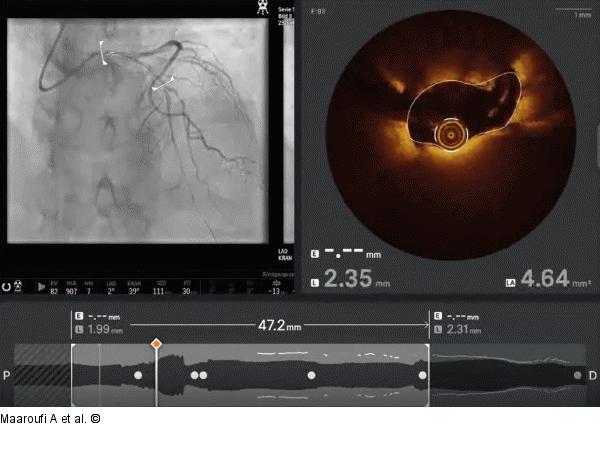
Abbildung 1: Video 1 Co-registration of OCT run in the LAD using Ultreon 2.0 software and Dragonfly Optis catheter. The video commences with the proximal segment of the left anterior descending artery (LAD) displaying simultaneous coronary angiogram on left and OCT imaging on right. Notably, the filling of the proximal LAD with contrast appears suboptimal in the angiogram, concomitant with the OCT images which vividly depict blood entering the vessel lumen. As the video progresses distally, the OCT frames exhibit significant artifact clearance from blood and better resolution of the vessel wall segments, concurrent with adequate contrast filling in the angiogram. This is a nice example how artificial intelligence (AI) can prepare imaging data for quick and easy review by the interventional cardiologist: In reality, blood is cleared from the lumen by antegrade dye injection and the OCT-lens is moved from distal to proximal, so in this case it reached the LM when blood already followed the contrast-bolus. AI used the coregistered images and presents the video in antegrade mode for more intuitive interpretation. Keep in mind, though, how the pictures were created, to better understand the creation of artifacts. In this case, less experienced cardiologists may misinterprete blood for thrombus if they only look at still frame OCT pictures. Co-registration of angio and OCT clearly shows the movement of blood. “It is all about the flow! |
Film starten 
Abbildung 1: Video 1
Co-registration of OCT run in the LAD using Ultreon 2.0 software and Dragonfly Optis catheter. The video commences with the proximal segment of the left anterior descending artery (LAD) displaying simultaneous coronary angiogram on left and OCT imaging on right. Notably, the filling of the proximal LAD with contrast appears suboptimal in the angiogram, concomitant with the OCT images which vividly depict blood entering the vessel lumen. As the video progresses distally, the OCT frames exhibit significant artifact clearance from blood and better resolution of the vessel wall segments, concurrent with adequate contrast filling in the angiogram. This is a nice example how artificial intelligence (AI) can prepare imaging data for quick and easy review by the interventional cardiologist: In reality, blood is cleared from the lumen by antegrade dye injection and the OCT-lens is moved from distal to proximal, so in this case it reached the LM when blood already followed the contrast-bolus. AI used the coregistered images and presents the video in antegrade mode for more intuitive interpretation. Keep in mind, though, how the pictures were created, to better understand the creation of artifacts. In this case, less experienced cardiologists may misinterprete blood for thrombus if they only look at still frame OCT pictures. Co-registration of angio and OCT clearly shows the movement of blood. “It is all about the flow! |


