Maaroufi A, Andreka J, Zirlik A, Toth GG, Harb S OCT-Corner: It's all about the flow Journal für Kardiologie - Austrian Journal of Cardiology 2024; 31 (7-8): 183-185 Volltext (PDF) Übersicht
| ||||||||||
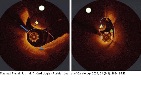
Abbildung 1: OCT Left: The imaging displays a reduction of the lumen due to an intra luminal protrusion of a high signal formation (yellow star) with high attenuation beneath it (white circle), intima (high signal) is intact and thin (blue triangles), the arrow indicates the coronary wire. Right: The high attenuation of signal beneath a protruding formation with a smooth surface, its proximal localization and intraluminal swirling contouring the wire (green triangle) in the following sequence of images, are all arguments suggesting an artifact related to the blood flowing into the coronary artery when flushing stopped before the pullback was ended. |

Abbildung 1: OCT
Left: The imaging displays a reduction of the lumen due to an intra luminal protrusion of a high signal formation (yellow star) with high attenuation beneath it (white circle), intima (high signal) is intact and thin (blue triangles), the arrow indicates the coronary wire. Right: The high attenuation of signal beneath a protruding formation with a smooth surface, its proximal localization and intraluminal swirling contouring the wire (green triangle) in the following sequence of images, are all arguments suggesting an artifact related to the blood flowing into the coronary artery when flushing stopped before the pullback was ended. |


