Smith ELK, Fan T-PD, Hainsworth AH, Hiley CR, Lauder H Vascular endothelial growth factor (VEGF)and endothelin-3 (ET-3) activate cation currents in human umbilical vein endothelial cells Journal of Clinical and Basic Cardiology 1998; 1 (1): 52-54 PDF Summary Overview
| ||||||
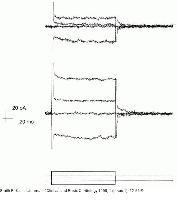
Figure/Graphic 1: VEGV und ET-3 - Gefäßpermeabilität Effect of acute exposure to vascular endothelial growth factor (VEGF) on whole cell currents recorded from human umbilical vein endothelial cells (HUVEC) on the edge of a regrowing boundary. Whole cell currents before (upper traces) and at the end of 10 min in VEGF (20 pmol/L) containing solution (middle traces). From a holding potential of -60mV, brief pulses to -100, -60, -20 and +40mV were applied (pulse protocol shown in bottom trace). Whole cell capacitance 24 pF, series resistance 5.5 MΩ. Currents are shown with residual capacity transients manually erased. Cell 601252. |

Figure/Graphic 1: VEGV und ET-3 - Gefäßpermeabilität
Effect of acute exposure to vascular endothelial growth factor (VEGF) on whole cell currents recorded from human umbilical vein endothelial cells (HUVEC) on the edge of a regrowing boundary. Whole cell currents before (upper traces) and at the end of 10 min in VEGF (20 pmol/L) containing solution (middle traces). From a holding potential of -60mV, brief pulses to -100, -60, -20 and +40mV were applied (pulse protocol shown in bottom trace). Whole cell capacitance 24 pF, series resistance 5.5 MΩ. Currents are shown with residual capacity transients manually erased. Cell 601252. |


