Content-type: text/html Suchergebnis - Abbildungen und Graphiken
Krause und Pachernegg
Verlag für Medizin und Wirtschaft
Mobile Version
A-  |   A  |   A+
Werbung
 
Suchergebnis - Abbildungen und Graphiken
 
Alle Journale wurden nach Gauster M durchsucht Treffer 1 - 3 von 3

Medizinische Publikationen (1)  ::   Abbildungen und Graphiken  ::   Volltext (0)

Neue Suche:         Erweiterte Suche    Hilfe zur Suche

in allen Journalen nach  Bildtitel    Bildkeywords   Bildsummary   Autoren      Mindestens ein Wort  Alle Wörter   
1. Gauster M; Huppertz B
Fusion of Cytothrophoblast with Syncytiotrophoblast in the Human Placenta: Factors Involved in Syncytialization
Trophoblast fusion
Journal für Reproduktionsmedizin und Endokrinologie - Journal of Reproductive Medicine and Endocrinology 2008; 5 (2): 76-82
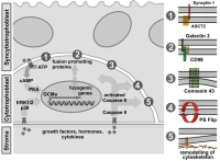
Illustration of factors involved in trophoblast fusion. Environmentally derived growthfactors, hormones and cytokines bind to their cognate receptors at the plasma membrane ofcytotrophoblasts. Activation of either protein kinase A (PKA) or MAP kinases ERK1/2 and p38leads to increased protein expression of the transcription factor GCMa, which in turn drivestranscription of fusogenic genes. Several structural and membrane proteins were suggested topromote trophoblast fusion, including syncytin 1 and its receptor ASCT2 (1), CD98 and its receptorgalectin 3 (2) as well as connexin 43 (3). Beside activation of PKA or MAP kinase pathways,cytokines induce conversion of pro-caspase 8 into active caspase 8. Once activated, caspase 8can mediate inactivation of “flippases†and/or activation of "floppases" to trigger phosphatidyl-serine externalisation (PS flip) (4). Additionally, caspase 8 triggers remodelling of the sub-mem-branous cytoskeleton by degrading structural proteins such as α-fodrin (5).

Keywords: Throphoblasttrophoblast fusion
2. Gauster M; Huppertz B
Fusion of Cytothrophoblast with Syncytiotrophoblast in the Human Placenta: Factors Involved in Syncytialization
Human placenta
Journal für Reproduktionsmedizin und Endokrinologie - Journal of Reproductive Medicine and Endocrinology 2008; 5 (2): 76-82
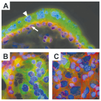
A) Immunofluorescent staining ofhuman first trimester placenta (week 8).Mononucleated cytotrophoblasts (arrow)show staining for E-cadherin in their apicaland lateral cell membranes (red). The over-lying multinucleated syncytiotrophoblast(arrowhead) lacks lateral cell walls and isnegative for E-cadherin. The syncytiotro-phoblast expresses hCG (green). B) and C)Immunofluorescent staining of forskolin andvehicle treated BeWo cells. Treatment ofBeWo cells for 48h with forskolin (20 μM)induced formation of multinucleated syn-cytia, visualized by degradation of E-cadherin (stained in red). hCG expression(green) was predominantly observed in syn-cytia of forskolin treated BeWo cells, butwas also rarely seen in mononucleatedBeWo cells (treated and untreated).

Keywords: placentaPlazenta
3. Gauster M; Huppertz B
Fusion of Cytothrophoblast with Syncytiotrophoblast in the Human Placenta: Factors Involved in Syncytialization
Trophoblast
Journal für Reproduktionsmedizin und Endokrinologie - Journal of Reproductive Medicine and Endocrinology 2008; 5 (2): 76-82
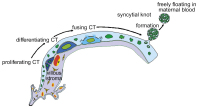
Schematic representation of trophoblast differentiation within the villous trophoblast.Derived from trophoblast progenitor cells daughter cells start to differentiate and finally fusewith the overlying syncytiotrophoblast. Nuclei that have become an integrative part of the syn-cytiotrophoblast change their morphology and display chromatin condensation. Finally lateapoptotic syncytial nuclei are packed into syncytial knots and are released into the maternalblood stream. The arrows follow the route of cell types and nuclear changes during trophoblastdifferentiation.

Keywords: SchemaschemeTrohoblasttrophoblast
 

 Seite 1 von 1 
 
copyright © 2000–2026 Krause & Pachernegg GmbH | Sitemap | Datenschutz | Impressum
 
Werbung