2.
Perneczky G;
Loyoddin M;
Schappelwein H;
Sherif C
Spinal Meningiomas: A Comprehensive Overview and Own Experience
Spinal Meningioma
European Association of NeuroOncology Magazine 2013; 3 (3): 118-121
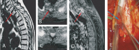
Pre- and intraoperative findings: (a) T2-weighted native sagittal MR image: the tumour can be nicely delineated (red arrow). (b, c) Axial T1-weighted contrastenhanced sequence: inhomogeneous contrast enhancement is due to intratumoural calcifications (red arrow). (d) Sagittal contrast-enhanced T1-weighted image: again, the calcifications can be seen as contrast agent-free areas within the tumour (red arrow). (e) Intraoperative findings: the spinal cord is carefully mobilized under IOM. The tumour (blue arrow) can be seen at the left ventral dura.
Keywords:
MRI,
spinal meningioma

 Suche Medizinische Publikationen
 Suche Abbildungen und Grafiken
 Medizinprodukte - Labortechnik
 Antidotarium (Stand 2012)
 Phytokodex (Stand 2012)
 Fachbücher
 Patientenliteratur