Content-type: text/html Suchergebnis - Abbildungen und Graphiken
Krause und Pachernegg
Verlag für Medizin und Wirtschaft
Mobile Version
A-  |   A  |   A+
Werbung
 
Suchergebnis - Abbildungen und Graphiken
 
Alle Journale wurden nach Polisca P durchsucht Treffer 1 - 4 von 4

Medizinische Publikationen (1)  ::   Abbildungen und Graphiken  ::   Volltext (0)

Neue Suche:         Erweiterte Suche    Hilfe zur Suche

in allen Journalen nach  Bildtitel    Bildkeywords   Bildsummary   Autoren      Mindestens ein Wort  Alle Wörter   
1. Gaspardone A; Chiariello L; Crea F; De Matteis GM; Gioffrè PA; Polisca P; Proietti I; Tomai F; Versaci F
Temporal Sequence and Spatial Distribution of Ischaemic Changes During Dipyridamole Stress Test - the Key Role of Microvascular Dysfunction
Different ischaemic events
Journal of Clinical and Basic Cardiology 2005; 8 (1-4): 47-53
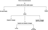
Flow chart showing the different ischaemic events in the study group during dipyridamole infusion after coronary artery revascularisation. Numbers indicate the number of patients. ECG: electrocardiographic changes; ECHO: echocardiographic changes; LAD-dep: systolic changes in left anterior descending coronary artery-dependent territories; non-LAD-dep: systolic changes in left anterior descending coronary artery-independent territories; LAD-dep and non-LAD-dep: systolic changes in left anterior descending coronary artery-dependent and -independent territories.

Keywords: ischaemiaIschämierevascularisationRevaskularisationSchemascheme
2. Gaspardone A; Chiariello L; Crea F; De Matteis GM; Gioffrè PA; Polisca P; Proietti I; Tomai F; Versaci F
Temporal Sequence and Spatial Distribution of Ischaemic Changes During Dipyridamole Stress Test - the Key Role of Microvascular Dysfunction
Different ischaemic events
Journal of Clinical and Basic Cardiology 2005; 8 (1-4): 47-53
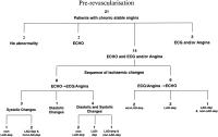
Flow chart showing the different ischaemic events in the study group during dipyridamole infusion before coronary artery revascularisation. Numbers indicate the number of patients. ECG: electrocardiographic changes; ECHO: echocardiographic changes; LAD-dep: systolic changes in left anterior descending coronary artery-dependent territories; non-LAD-dep: systolic changes in left anterior descending coronary artery independent territories; LAD-dep and non-LAD-dep: systolic changes in left anterior descending coronary artery-dependent and independent territories.

Keywords: ischaemiaIschämieSchemascheme
3. Gaspardone A; Chiariello L; Crea F; De Matteis GM; Gioffrè PA; Polisca P; Proietti I; Tomai F; Versaci F
Temporal Sequence and Spatial Distribution of Ischaemic Changes During Dipyridamole Stress Test - the Key Role of Microvascular Dysfunction
Ischaemic events - Temporal sequence
Journal of Clinical and Basic Cardiology 2005; 8 (1-4): 47-53
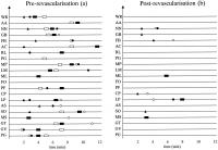
Schematic representation of the temporal sequence of ischaemic events during dipyridamole infusion in each patient before (panel a) and after (panel b) coronary artery revascularisation. Black squares: angina pectoris; white squares: electrocardiographic ischaemic changes; black circles: echocardiographic alterations in non-LAD-dependent myocardial segments; white circles: echocardiographic alterations in LAD-dependent myocardial segments; black triangles: Doppler mitral flow abnormality. LAD: left anterior descending coronary artery

Keywords: ischaemiaIschämieSchemascheme
4. Gaspardone A; Chiariello L; Crea F; De Matteis GM; Gioffrè PA; Polisca P; Proietti I; Tomai F; Versaci F
Temporal Sequence and Spatial Distribution of Ischaemic Changes During Dipyridamole Stress Test - the Key Role of Microvascular Dysfunction
Wall motion analysis
Journal of Clinical and Basic Cardiology 2005; 8 (1-4): 47-53
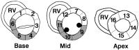
Sixteen-segment model for wall motion analysis. White segments were considered LAD-dependent myocardial segments (n = 10); shaded segments were considered non-LAD-dependent myocardial segments (n = 6). 1: proximal anterior septum; 2: proximal anterior wall; 3: proximal lateral wall; 4: proximal posterior wall; 5: proximal inferior wall; 6: proximal inferior septum; 7: mid anterior wall; 8: mid lateral wall; 9: mid posterior wall; 10: mid inferior wall; 11: mid inferior septum; 12: mid anterior septum; 13: distal anterior wall; 14: distal lateral wall; 15: distal posterior wall; 16: distal anterior septum. RV: right ventricle

Keywords: LADSchemaschemewall motion analysis
 

 Seite 1 von 1 
 
copyright © 2000–2026 Krause & Pachernegg GmbH | Sitemap | Datenschutz | Impressum
 
Werbung