1.
Kuntz-Hehner St;
Becher H;
Luederitz B;
Omran H;
Schlosser Th;
Tiemann K
Assessment of Myocardial Perfusion by Contrast Echocardiography - Ready for Clinical Practice?
Kontrastechokardiographie
Journal of Clinical and Basic Cardiology 2002; 5 (2): 145-148
Negative bolus de
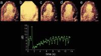
struction reperfusion study using power pulse inversion imaging. First, continuous infusion of a contrast agent is adjusted to achieve full myocardial opacification using real-time imaging at very low emission power (a) (MI = 0.09). After destruction of the microbubbles by a short period of high power imaging (b) (MI = 1.3) myocardial contrast replenishment is recorded at low emission power (c–e) (MI = 0.09). A region of interest (ROI) is placed in the septum and repeated
measurements are made at the endsystolic frames. The graph shows the reperfusion curve for this ROI, fitting well to the monoexponential function.
Keywords:
Contrast agent,
echocardiography,
Echokardiographie,
Kontrastmittel,
reperfusion,
reperfusion

 Suche Medizinische Publikationen
 Suche Abbildungen und Grafiken
 Medizinprodukte - Labortechnik
 Antidotarium (Stand 2012)
 Phytokodex (Stand 2012)
 Fachbücher
 Patientenliteratur