Krause und Pachernegg
Verlag für Medizin und Wirtschaft
Artikel   Bilder   Volltext

Mobile Version
A-  |   A  |   A+
Werbung
 
Figures and Graphics
Proescholdt M et al.  
Surgery of Malignant Gliomas Using Modern Technology

European Association of NeuroOncology Magazine 2013; 3 (1): 11-14

PDF    Summary    Figures   


Figure
 
Glioma - Surgery
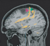
Figure 1: Fusion of [F-18]fluoroethyltyrosine: PET (red), functional MRI (green), and DTI tractography (yellow) in a patient with a left frontal anaplastic astrocytoma. Note the close vicinity of eloquent cortical and subcortical structures to the tumour borders.


Keywords: gliomaMRIPET
 
 
Glioma - Surgery
Figure 2a-c: Quantification of the preoperative tumour volume in malignant gliomas. (A) Axial T1-weighted MRI scan of a patient with a left frontal glioblastoma. (B) The contrast-enhancing part of each section is outlined and subsequently fused to generate (C) a 3-dimensional segment which can be quantified volumetrically.


Keywords: gliomaMRITumor


copyright © 2000–2025 Krause & Pachernegg GmbH | Sitemap | Datenschutz | Impressum