Proescholdt M, Doenitz C, Brawanski A Surgery of Malignant Gliomas Using Modern Technology European Association of NeuroOncology Magazine 2013; 3 (1): 11-14 PDF Summary Overview
| ||||
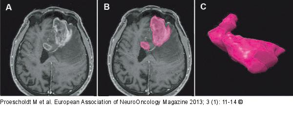
Figure/Graphic 2a-c: Glioma - Surgery Quantification of the preoperative tumour volume in malignant gliomas. (A) Axial T1-weighted MRI scan of a patient with a left frontal glioblastoma. (B) The contrast-enhancing part of each section is outlined and subsequently fused to generate (C) a 3-dimensional segment which can be quantified volumetrically. |

Figure/Graphic 2a-c: Glioma - Surgery
Quantification of the preoperative tumour volume in malignant gliomas. (A) Axial T1-weighted MRI scan of a patient with a left frontal glioblastoma. (B) The contrast-enhancing part of each section is outlined and subsequently fused to generate (C) a 3-dimensional segment which can be quantified volumetrically. |


