| Rosenhek R, Binder T |
|---|
Monitoring of Invasive Procedures - The Role of Echocardiography in Cathlab and Operating Room
Journal of Clinical and Basic Cardiology 2002; 5 (2): 139-143
PDF Summary Figures
| Figure |
|---|
| |
|
|
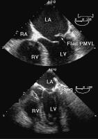
Echokardiographie - Mitralklappen-OP
Figure 1: Flail posterior mitral valve leaflet (PMVL) as mechanism of mitral regurgitation (top panel). Immediate postoperative result after mitral valve reconstruction with annuloplasty ring implantation (lower panel). LA = left atrium, LV = left ventricle, RV = right ventricle
Keywords: Annuloplastie,
annuloplasty,
echocardiography,
Echokardiographie,
Mitral valve,
Mitralklappe,
Reconstruction,
Rekonstruktion
|
| |
| |
|
|
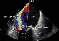
Echokardiographie - Mitralklappe
Figure 2: Large paravalvular leak detected by color Doppler echocardiography in a bioprosthetic mitral valve (MV-Bio Prosth). LA = left atrium, LV = left ventricle
Keywords: Colordoppler,
echocardiography,
Echokardiographie,
Farbdoppler,
Mitral valve,
Mitralklappe,
Prosthesis,
Prothese
|
| |
| |
|
|
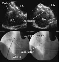
Amplatzer-Occluder - Positionierung
Figure 3: Positioning of an Amplatzer atrial septal defect occluder under TEE guidance (top left panel) and assessment of the result after release of the occluder (top right panel). The lower panels show the corresponding images obtained by fluoroscopy. LA = left atrium, RA = right atrium
Keywords: Amplatzer-Occluder,
Amplatzer-Occluder,
echocardiography,
Echokardiographie,
Fluoroscopy,
Fluoroskopie,
TEE,
TEE
|
| |
| |
|
|
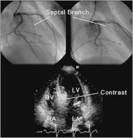
Echokardiographie - Kardiomyopathie
Figure 4: The septal branch of the left anterior descending coronary artery in a patient with hypertrophic obstructive cardiomyopathy (top panels). Estimation of the size of the septal vascular territory with myocardial contrast echocardiography (lower panel). LA = left atrium, RA = right atrium, LV = left ventricle, RV = right ventricle
Keywords: Angiographie,
Arteria coronaria sinistra,
Arteria coronaria sinistra,
cardiomyopathy,
echocardiography,
Echokardiographie,
Kardiomyopathie
|
| |
| |
|
|


