Rosenhek R, Binder T Monitoring of Invasive Procedures - The Role of Echocardiography in Cathlab and Operating Room Journal of Clinical and Basic Cardiology 2002; 5 (2): 139-143 PDF Summary Overview
| ||||||||
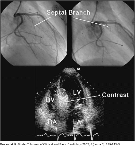
Figure/Graphic 4: Echokardiographie - Kardiomyopathie The septal branch of the left anterior descending coronary artery in a patient with hypertrophic obstructive cardiomyopathy (top panels). Estimation of the size of the septal vascular territory with myocardial contrast echocardiography (lower panel). LA = left atrium, RA = right atrium, LV = left ventricle, RV = right ventricle |

Figure/Graphic 4: Echokardiographie - Kardiomyopathie
The septal branch of the left anterior descending coronary artery in a patient with hypertrophic obstructive cardiomyopathy (top panels). Estimation of the size of the septal vascular territory with myocardial contrast echocardiography (lower panel). LA = left atrium, RA = right atrium, LV = left ventricle, RV = right ventricle |


