| Grossman R, Ram Z | ||||||||||||||||||||||||||||||||||
|---|---|---|---|---|---|---|---|---|---|---|---|---|---|---|---|---|---|---|---|---|---|---|---|---|---|---|---|---|---|---|---|---|---|---|
|
Awake Craniotomy in Glioma Surgery
European Association of NeuroOncology Magazine 2014; 4 (1): 27-33 PDF Summary Figures
|
||||||||||||||||||||||||||||||||||

Verlag für Medizin und Wirtschaft |
|
||||||||
|
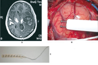
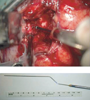
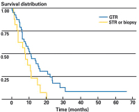
Figures and Graphics
|
|||||||||
| copyright © 2000–2025 Krause & Pachernegg GmbH | Sitemap | Datenschutz | Impressum | |||||||||


