Grossman R, Ram Z Awake Craniotomy in Glioma Surgery European Association of NeuroOncology Magazine 2014; 4 (1): 27-33 PDF Summary Overview
| ||||||||||
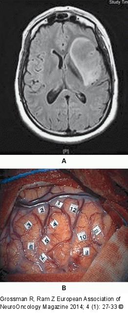
Figure/Graphic 2A-B: Awake Craniotomy (A) Pre-operative axial fluid-attenuated inversion recovery- (FLAIR-) weighted MRI showing a WHO grade-II insular glioma in a 40-year-old woman presenting with episodes of seizures. This patient has no neurological deficit. (B) Intra-operative cortical mapping of language during awake surgery. |

Figure/Graphic 2A-B: Awake Craniotomy
(A) Pre-operative axial fluid-attenuated inversion recovery- (FLAIR-) weighted MRI showing a WHO grade-II insular glioma in a 40-year-old woman presenting with episodes of seizures. This patient has no neurological deficit. (B) Intra-operative cortical mapping of language during awake surgery. |


