Grossman R, Ram Z Awake Craniotomy in Glioma Surgery European Association of NeuroOncology Magazine 2014; 4 (1): 27-33 PDF Summary Overview
| ||||||||||
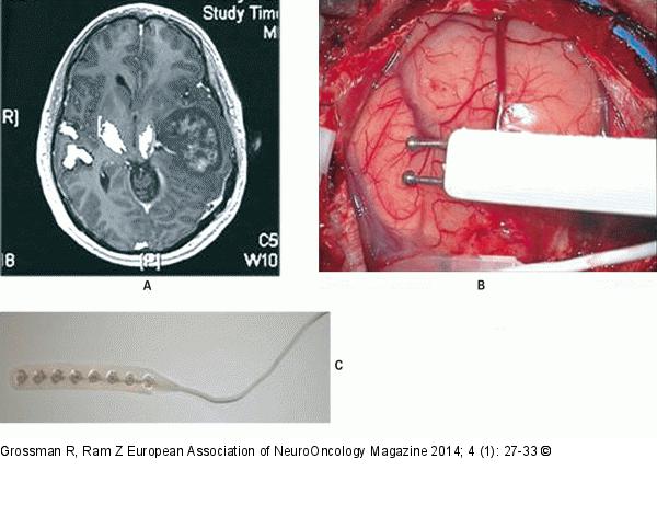
Figure/Graphic 3A-C: Awake Craniotomy (A) Preoperative T1-weighted axial after gadolinium injection with diffuse tensor imaging (DTI) of the cortico-spinal tract showing a left temporo-parietal high-grade glioma displace medially the corticospinal tract. (B) Direct cortical stimulation by using bipolar probe. (C) A cortical strip electrode is placed over the surface of the motor cortex in order to assess motor-evoked potential (MEP). |

Figure/Graphic 3A-C: Awake Craniotomy
(A) Preoperative T1-weighted axial after gadolinium injection with diffuse tensor imaging (DTI) of the cortico-spinal tract showing a left temporo-parietal high-grade glioma displace medially the corticospinal tract. (B) Direct cortical stimulation by using bipolar probe. (C) A cortical strip electrode is placed over the surface of the motor cortex in order to assess motor-evoked potential (MEP). |


