Kuntz-Hehner St, Becher H, Luederitz B, Omran H, Schlosser Th, Tiemann K Assessment of Myocardial Perfusion by Contrast Echocardiography - Ready for Clinical Practice? Journal of Clinical and Basic Cardiology 2002; 5 (2): 145-148 PDF Summary Overview
| ||||
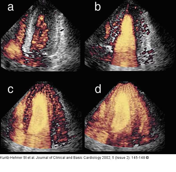
Figure/Graphic 1a-d: Kontrastechokardiographie Different degrees of myocardial contrast using harmonic power Doppler imaging (four chamber view) during continuous infusion of Levovist (R): Arrival of the contrast agent in the right ventricle (a) and complete left ventricular opacification shortly after (b) using triggered imaging every heart cycle (1:1). Complete left ventricular myocardial opacification using higher trigger intervals every 3rd (c) and every 5th (d) cardiac cycle. |

Figure/Graphic 1a-d: Kontrastechokardiographie
Different degrees of myocardial contrast using harmonic power Doppler imaging (four chamber view) during continuous infusion of Levovist (R): Arrival of the contrast agent in the right ventricle (a) and complete left ventricular opacification shortly after (b) using triggered imaging every heart cycle (1:1). Complete left ventricular myocardial opacification using higher trigger intervals every 3rd (c) and every 5th (d) cardiac cycle. |


