Pfabe FP EndoAnchor-Implantation: Stellenwert, Möglichkeiten und Grenzen bei EVAR und „hostile neck“-Anatomie // EndoAnchor – implantation: value, possibilities and limits at EVAR and “hostile neck” Zeitschrift für Gefäßmedizin 2019; 16 (3): 10-15 Volltext (PDF) Summary Übersicht
| ||||||||||||||||
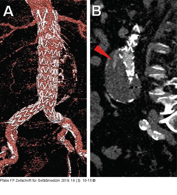
Abbildung 2: EVAR-Prozedur Verlaufskontrolle nach EVAR-Prozedur mittels CT-Angiographie nach 9 Monaten. (A): Darstellung in VRT, regelrechte Entfaltung der Prothese; (B): sagittale Schnittebene, Nachweis eines Endoleaks Typ Ia ventrolateral (roter Pfeil). |

Abbildung 2: EVAR-Prozedur
Verlaufskontrolle nach EVAR-Prozedur mittels CT-Angiographie nach 9 Monaten. (A): Darstellung in VRT, regelrechte Entfaltung der Prothese; (B): sagittale Schnittebene, Nachweis eines Endoleaks Typ Ia ventrolateral (roter Pfeil). |


