|
|
|---|
1. Schuchlenz H;
Pachler C;
Binder R;
Mair J;
Toth GG;
Gabriel H
Review und Leitlinien für die Diagnostik und den interventionellen Verschluss des persistierenden Foramen ovale (PFO) // Review and guidelines for diagnosis and interventional closure of PFO
TEE-Studie
Journal für Kardiologie - Austrian Journal of Cardiology 2019; 26 (7-8): 178-192
TEE-Studie mit Thrombus „in transit“. Keywords: Echo,
Kardiologie,
TEE,
Thrombus
|
|
|
2. Schuchlenz H;
Pachler C;
Binder R;
Mair J;
Toth GG;
Gabriel H
Review und Leitlinien für die Diagnostik und den interventionellen Verschluss des persistierenden Foramen ovale (PFO) // Review and guidelines for diagnosis and interventional closure of PFO
TEE-Schnittbilder
Journal für Kardiologie - Austrian Journal of Cardiology 2019; 26 (7-8): 178-192
TEE-Schnittbilder: Bikavaler Schnitt (a), kurze Achse (b), PFO-Anatomie (c).
© H. Schuchlenz Keywords: Kardiologie,
Schema,
TEE
|
|
|
3. Schuchlenz H;
Pachler C;
Binder R;
Mair J;
Toth GG;
Gabriel H
Review und Leitlinien für die Diagnostik und den interventionellen Verschluss des persistierenden Foramen ovale (PFO) // Review and guidelines for diagnosis and interventional closure of PFO
Fossa ovalis
Journal für Kardiologie - Austrian Journal of Cardiology 2019; 26 (7-8): 178-192
Fossa ovalis (FO) in der Frontalansicht
(a) und von inferior (b). © H.
Schuchlenz Keywords: Fossa ovalis,
Kardiologie,
Schema
|
|
|
4. Schuchlenz H;
Pachler C;
Binder R;
Mair J;
Toth GG;
Gabriel H
Review und Leitlinien für die Diagnostik und den interventionellen Verschluss des persistierenden Foramen ovale (PFO) // Review and guidelines for diagnosis and interventional closure of PFO
PFO
Journal für Kardiologie - Austrian Journal of Cardiology 2019; 26 (7-8): 178-192
PFO mit Kanallänge (a), Größe = Distanz zwischen
dünnem Septum primum und dickerem Septum secundum (b),
sowie Vorhofseptumaneurysma (c). © H. Schuchlenz Keywords: Kardiologie,
PFO,
Schema
|
|
|
5. Schuchlenz H;
Pachler C;
Binder R;
Mair J;
Toth GG;
Gabriel H
Review und Leitlinien für die Diagnostik und den interventionellen Verschluss des persistierenden Foramen ovale (PFO) // Review and guidelines for diagnosis and interventional closure of PFO
PFO
Journal für Kardiologie - Austrian Journal of Cardiology 2019; 26 (7-8): 178-192
PFO mit kleiner (a) und großer Valvula Eustachii (b) (Pfeil). Keywords: Echo,
Kardiologie,
PFO,
Valvula eustachii
|
|
|
6. Schuchlenz H;
Pachler C;
Binder R;
Mair J;
Toth GG;
Gabriel H
Review und Leitlinien für die Diagnostik und den interventionellen Verschluss des persistierenden Foramen ovale (PFO) // Review and guidelines for diagnosis and interventional closure of PFO
Vorhofseptumaneurysma
Journal für Kardiologie - Austrian Journal of Cardiology 2019; 26 (7-8): 178-192
Vorhofseptumaneurysma: TEE-Studie, bikavaler Schnitt, PFO mit Vorhofseptumaneurysma (Pfeil) Keywords: Echo,
Kardiologie,
Vorhofseptumaneurysma
|
|
|
7. Schuchlenz H;
Pachler C;
Binder R;
Mair J;
Toth GG;
Gabriel H
Review und Leitlinien für die Diagnostik und den interventionellen Verschluss des persistierenden Foramen ovale (PFO) // Review and guidelines for diagnosis and interventional closure of PFO
Vorhofseptum
Journal für Kardiologie - Austrian Journal of Cardiology 2019; 26 (7-8): 178-192
Entwicklung des Vorhofseptums, Septum primum
linksatrial: rot, Septum secundum rechtsatrial: blau (siehe Text).
© H. Schuchlenz Keywords: Kardiologie,
Schema,
Vorhofseptum
|
|
|
8. Rosenhek R;
Binder T
Monitoring of Invasive Procedures - The Role of Echocardiography in Cathlab and Operating Room
Echokardiographie - Kardiomyopathie
Journal of Clinical and Basic Cardiology 2002; 5 (2): 139-143
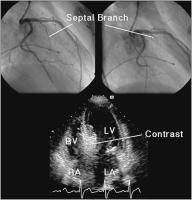
The septal branch of the left anterior descending coronary artery in a patient with hypertrophic obstructive cardiomyopathy (top panels). Estimation of the size of the septal vascular territory with myocardial contrast echocardiography (lower panel). LA = left atrium, RA = right atrium, LV = left ventricle, RV = right ventricle Keywords: Angiographie,
Arteria coronaria sinistra,
Arteria coronaria sinistra,
cardiomyopathy,
echocardiography,
Echokardiographie,
Kardiomyopathie
|
|
|
9. Rosenhek R;
Binder T
Monitoring of Invasive Procedures - The Role of Echocardiography in Cathlab and Operating Room
Amplatzer-Occluder - Positionierung
Journal of Clinical and Basic Cardiology 2002; 5 (2): 139-143
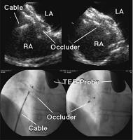
Positioning of an Amplatzer atrial septal defect occluder under TEE guidance (top left panel) and assessment of the result after release of the occluder (top right panel). The lower panels show the corresponding images obtained by fluoroscopy. LA = left atrium, RA = right atrium Keywords: Amplatzer-Occluder,
Amplatzer-Occluder,
echocardiography,
Echokardiographie,
Fluoroscopy,
Fluoroskopie,
TEE,
TEE
|
|
|
10. Rosenhek R;
Binder T
Monitoring of Invasive Procedures - The Role of Echocardiography in Cathlab and Operating Room
Echokardiographie - Mitralklappe
Journal of Clinical and Basic Cardiology 2002; 5 (2): 139-143
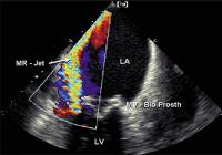
Large paravalvular leak detected by color Doppler echocardiography in a bioprosthetic mitral valve (MV-Bio Prosth). LA = left atrium, LV = left ventricle Keywords: Colordoppler,
echocardiography,
Echokardiographie,
Farbdoppler,
Mitral valve,
Mitralklappe,
Prosthesis,
Prothese
|
|
|
11. Rosenhek R;
Binder T
Monitoring of Invasive Procedures - The Role of Echocardiography in Cathlab and Operating Room
Echokardiographie - Mitralklappen-OP
Journal of Clinical and Basic Cardiology 2002; 5 (2): 139-143
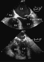
Flail posterior mitral valve leaflet (PMVL) as mechanism of mitral regurgitation (top panel). Immediate postoperative result after mitral valve reconstruction with annuloplasty ring implantation (lower panel). LA = left atrium, LV = left ventricle, RV = right ventricle Keywords: Annuloplastie,
annuloplasty,
echocardiography,
Echokardiographie,
Mitral valve,
Mitralklappe,
Reconstruction,
Rekonstruktion
|
|
|
| |

 Suche Medizinische Publikationen
 Suche Abbildungen und Grafiken
 Medizinprodukte - Labortechnik
 Antidotarium (Stand 2012)
 Phytokodex (Stand 2012)
 Fachbücher
 Patientenliteratur