Content-type: text/html Suchergebnis - Abbildungen und Graphiken
Krause und Pachernegg
Verlag für Medizin und Wirtschaft
Mobile Version
A-  |   A  |   A+
Werbung
 
Suchergebnis - Abbildungen und Graphiken
 
Alle Journale wurden nach Nicolosi GL durchsucht Treffer 1 - 4 von 4

Medizinische Publikationen (1)  ::   Abbildungen und Graphiken  ::   Volltext (0)

Neue Suche:         Erweiterte Suche    Hilfe zur Suche

in allen Journalen nach  Bildtitel    Bildkeywords   Bildsummary   Autoren      Mindestens ein Wort  Alle Wörter   
1. Nicolosi GL
Infective Endocarditis: Diagnostic and Therapeutic Issues - Should Transesophageal Echocardiography be Performed in all Patients? - Position Con
Endokarditis - Diagnose - TEE
Journal of Clinical and Basic Cardiology 2001; 4 (2): 161-164
Two short axis transoesophageal views, 15 days apart, from an elderly patient with calcific aortic valve and infective endocarditis. In the first examination (top) a vegetation could not be identified since the echogenic nodularities were indistinguishable from fibrocalcific deposits (arrow). In the second examination (bottom) a vegetation was diagnosed on the aortic valve for the evident increase in size of one of the nodularities (arrow) which was still not mobile in real time (see text). A= aortic valve; LA= left atrium

Keywords: echocardiographyEchokardiographieendocarditisEndokarditis
2. Nicolosi GL
Infective Endocarditis: Diagnostic and Therapeutic Issues - Should Transesophageal Echocardiography be Performed in all Patients? - Position Con
Endokarditis - Diagnose - TEE
Journal of Clinical and Basic Cardiology 2001; 4 (2): 161-164
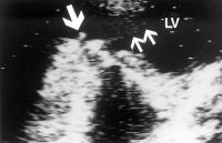
Transthoracic modified 4 chamber apical view in a patient with a mitral mechanical prosthetic valve, where the disease was only evident on the ventricular side of the valve itself (large arrow), which was masking its presence by the transoesophageal approach. The two small arrows indicate a spontaneous echo contrast small diastolic jet through the stenotic prosthetic valve, due to ingrowing tissue (see text). LV = left ventricle

Keywords: echocardiographyEchokardiographieendocarditisEndokarditis
3. Nicolosi GL
Infective Endocarditis: Diagnostic and Therapeutic Issues - Should Transesophageal Echocardiography be Performed in all Patients? - Position Con
Endokarditis - Diagnose - TEE
Journal of Clinical and Basic Cardiology 2001; 4 (2): 161-164
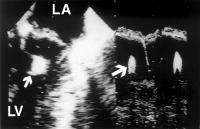
Systolic frame from a 4 chamber transesophageal echocardiographic study in a patient with chronic renal failure and haemodyalisis. The unexpected oscillating mass on the submitral apparatus (arrows: left 2D, right M-mode derived echocardiogram) is consistent with vegetation (see text). LA = left atrium; LV = left ventricle.

Keywords: echocardiographyEchokardiographieendocarditisEndokarditis
4. Nicolosi GL
Infective Endocarditis: Diagnostic and Therapeutic Issues - Should Transesophageal Echocardiography be Performed in all Patients? - Position Con
Endokarditis - Diagnose - TEE
Journal of Clinical and Basic Cardiology 2001; 4 (2): 161-164
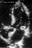
Apical transthoracic 5 chamber view from a 59 year old male with fever, non-Hodgkin lymphoma and an echogenic mass, mobile in real time, at the base of the posterior leaflet of the mitral valve (m). This could be classified as a positive echocardiogram and a major Duke criteria for the diagnosis of IE (see text)

Keywords: echocardiographyEchokardiographieendocarditisEndokarditis
 

 Seite 1 von 1 
 
copyright © 2000–2026 Krause & Pachernegg GmbH | Sitemap | Datenschutz | Impressum
 
Werbung