Content-type: text/html Suchergebnis - Abbildungen und Graphiken
Krause und Pachernegg
Verlag für Medizin und Wirtschaft
Mobile Version
A-  |   A  |   A+
Werbung
 
Suchergebnis - Abbildungen und Graphiken
 
Alle Journale wurden nach Schackert G durchsucht Treffer 1 - 7 von 7

Medizinische Publikationen (3)  ::   Abbildungen und Graphiken  ::   Volltext (0)

Neue Suche:         Erweiterte Suche    Hilfe zur Suche

in allen Journalen nach  Bildtitel    Bildkeywords   Bildsummary   Autoren      Mindestens ein Wort  Alle Wörter   
1. Uckermann O; Galli R; Mackenroth L; Geiger K; Steiner G; Koch E; Schackert G; Kirsch M
Optical Biochemical Imaging: Potential New Applications in Neuro-Oncology
NLO imaging
European Association of NeuroOncology Magazine 2014; 4 (1): 20-26
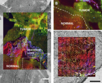
Multimodal NLO imaging of human tumours (red: CARS, green: TPEF, blue: SHG), overlaid with the bright field images of the unstained sample. The technique allows to retrieve detailed morphochemical information about tissue structure and properties on unstained samples. (a) Cryosection of human glioblastoma (scale bar: 200 μm). (b) High magnification of the border between tumour and normal tissue as indicated in (a). (c) Cryosection of human neuroma (scale bar: 0.5 mm).

Keywords: NLO imagingnonlinear optical microscopy
2. Uckermann O; Galli R; Mackenroth L; Geiger K; Steiner G; Koch E; Schackert G; Kirsch M
Optical Biochemical Imaging: Potential New Applications in Neuro-Oncology
Multimodal NLO
European Association of NeuroOncology Magazine 2014; 4 (1): 20-26
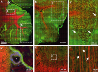
Multimodal NLO imaging of a mouse cerebellum (red: CARS, green: TPEF, blue: SHG). (a) Overview of the mouse cerebellum. (b) Magnification of the area indicated in (a). (c) Magnification of the area indicated in (b). Arrows indicate Purkinje cells characterized by punctuate fluorescence in the somata. (d) Magnification of the area indicated in (a), a large blood vessel is shown (*). (e) Magnification of the area indicated in (a), nerve fibres are indicated by intense CARS signal. (f) Magnification of the area indicated in (e). Single axons can be discerned (arrows).

Keywords: NLO imagingnonlinear optical microscopy
3. Uckermann O; Galli R; Mackenroth L; Geiger K; Steiner G; Koch E; Schackert G; Kirsch M
Optical Biochemical Imaging: Potential New Applications in Neuro-Oncology
CARS image
European Association of NeuroOncology Magazine 2014; 4 (1): 20-26
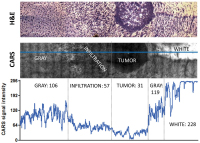
CARS microscopy of infiltrative experimental glioblastoma in a mouse model: The intensity of the CARS image and the morphological details permit to distinguish between the tumour mass, the infiltrative area, and the normal brain grey and white matters. H&E staining (top) CARS image (centre). The intensity of the CARS signal along the blue line is plotted in the graph (bottom). Mean intensity values for the different types of tissue are also reported. Mean intensity that characterizes the normal grey tissue is reduced to approximately 50 % in the infiltrative area and to approximately 30 % in the tumour. Lipid rich white matter is characterized by high CARS signal intensity (228, saturation in some areas).

Keywords: CARS imagescheme
4. Uckermann O; Galli R; Mackenroth L; Geiger K; Steiner G; Koch E; Schackert G; Kirsch M
Optical Biochemical Imaging: Potential New Applications in Neuro-Oncology
FT-IR imaging
European Association of NeuroOncology Magazine 2014; 4 (1): 20-26
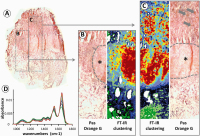
FT-IR imaging of human pituitary adenoma. (a) Overview of a cryosection of human pituitary adenoma stained using Pas-Orange-G and illustrating the production of human growth hormone (HGH). (b, c) High magnification of the areas indicated in (a) and corresponding FT-IR spectroscopic images obtained by cluster analysis. Large areas with increased HGH production are indicated by asterisks, local spots of hormone production are marked by arrows. (d) Representative spectra corresponding to area (b).

Keywords: FT-IR imaginghuman pituitary adenomascheme
5. Uckermann O; Galli R; Mackenroth L; Geiger K; Steiner G; Koch E; Schackert G; Kirsch M
Optical Biochemical Imaging: Potential New Applications in Neuro-Oncology
Vibrational Spectroscopy
European Association of NeuroOncology Magazine 2014; 4 (1): 20-26
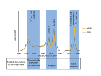
FT-IR spectra of grey and white matters of nervous tissue. The main bond vibrations and related dominating tissue components are indicated.

Keywords: FT-IR spectraschemevibrational spectroscopy
6. Krex D; Rudolph K; Schackert G
Die Behandlung von älteren Patienten mit benignen oder malignen Hirntumoren
Glioblastom
Journal für Neurologie, Neurochirurgie und Psychiatrie 2011; 12 (2): 142-147
Anteile adjuvanter Therapien bei Glioblastompatienten.

Keywords: DiagrammGlioblastomNeurochirurgieTherapie
7. Krex D; Rudolph K; Schackert G
Die Behandlung von älteren Patienten mit benignen oder malignen Hirntumoren
Hirntumor - Alter
Journal für Neurologie, Neurochirurgie und Psychiatrie 2011; 12 (2): 142-147
Klinischer Zustand der Gesamtpopulation in den beiden Altersgruppen 75+ (a) und 50– (b). „Selbständig“ = „Karnofsky Performance Score“ (KPS) 80–100 %; „auf Hilfe angewiesen“ = KPS 60–70 %, „pflegebedürftig“ = KPS 40–50 %.

Keywords: AlterDiagrammHirntumorNeurochirurgie
 

 Seite 1 von 1 
 
copyright © 2000–2026 Krause & Pachernegg GmbH | Sitemap | Datenschutz | Impressum
 
Werbung