Krause und Pachernegg
Verlag für Medizin und Wirtschaft
Artikel   Bilder   Volltext

Mobile Version
A-  |   A  |   A+
Werbung
 
Abbildungen und Graphiken
Koyama K, Hasegawa A  
Premature Ovarian Failure Syndrome May Be Induced by Autoimmune Reactions to Zona Pellucida Proteins

Journal für Reproduktionsmedizin und Endokrinologie - Journal of Reproductive Medicine and Endocrinology 2006; 3 (2): 94-97

Volltext (PDF)    Summary    Abbildungen   


Abbildung
 
Zona pellucida
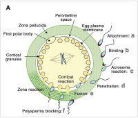
Abbildung 1a: Role of the zona pellucida (ZP) in fertilisation and oocyte maturation. A: Schematic drawing of sperm-ZP interaction (modified from [11]). The capacitated sperm attaches and then tightly binds to the ZP (a, b). These interactions are mediated by ZP3 which serves as a primary receptor for sperm. The acrosome reaction in the bound sperm is induced which is essential for sperm penetration of ZP (c). ZP2 maintains acrosomereacted sperm in the ZP during penetration (d). Following fusion of egg and sperm, cortical granules disintegrate to induce the zona reaction that prevents polyspermy (e, f). Proteolytic cleavage of ZP2 and ZP3 is involved in the zona reaction. After fertilisation ZP still remains to protect the fertilised eggs.


Keywords: Fertilisationoocyte maturationOozyteSchemaschemeZona pellucida
 
 
Zona pellucida
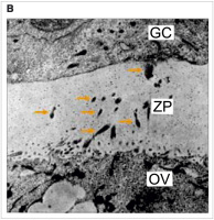
Abbildung 1b: Electron microscopic picture of a mouse oocyte section, demonstrating the zona pellucida (ZP), framed by a granulosa cell (GC) and the oocyte (OV). Protruding micro-villi of the granulosa cell are visible within the ZP.


Keywords: electron microscopic pictureElektronenmikroskopoocyteOozyteZona pellucida
 
 
Anti-Zona pellucida antibodies
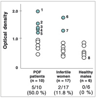
Abbildung 2: Detection of anti-ZP antibodies in POF patients by dot immunoassay. The human zona pellucida was isolated from unfertilised oocytes in a clinical program of IVF-ET under informed consent. Solubilised protein (0.01 µg) was dotted onto a nitrocellulose membrane. The membrane was incubated with the patients’ diluted sera and subsequently treated with an anti-human IgG antibody conjugated with horseradish peroxidase. Color development was performed by chloronaphthol and H2O2. Densitometric values showing more than twice over the SD values of the control group (healthy males) were assessed as positive. 50 % (5/10) of POF patients showed positive reaction for anti-zona antibodies, while 11.8 % (2/17) were positive in the other infertile women.


Keywords: antibodyAntikörperDiagrammflowchartPOFpremature ovarian failureZona pellucida
 
 
POF - oocytes
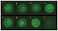
Abbildung 3: Immunofluorescent staining of human oocytes with sera from POF and the other infertile patients. Isolated human oocytes prepared as described in Fig. 2 were treated with patients’ diluted sera for one hour at room temperature. After washing with PBS, the treated oocytes were further incubated in FITC-conjugated anti-human IgG for 50 minutes. Observations were carried out under a fluorescent microscope. Each number corresponds to the serum samples in Fig. 2. The immunofluorescent intensities did not correlate to the values in the dot immunoassay. (1)–(5) are oocytes stained with POF patients’ sera, (6) and (7) are oocytes stained with other infertile patients’ sera. (8) is an oocyte stained with a healthy male’s serum showing a negative reaction in dot immunoassay.


Keywords: oocyteOozytePOFpremature ovarian failure
 
 
Antibodies - ELISA
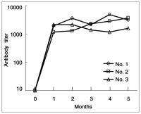
Abbildung 4: Increase of antibody titers in ELISA against the synthetic peptide used for immunisation. An 18-mer synthetic peptide of dog ZP2 (88–106 amino acids) was prepared and conjugated with a carrier protein, diphtheria toxoid for immunisation. The immunogen (500 µg) was injected with complete Freund’s adjuvant into three 3-month-old dogs at one-month intervals. Antibody titers were determined by ELISA against the cognate peptide antigen.


Keywords: antibodyAntikörperDiagrammELISAflowchart
 
 
ZP2 peptide
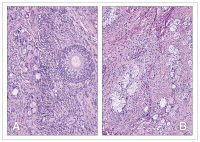
Abbildung 5: Histological examination of ovarian sections from dog immunised with ZP2 peptide. As compared to the control (A), the ovarian section from the immunised dog showed remarkable impairment (B). No growing follicle was detected and a fibrous change was observed in the interstitial tissue.


Keywords: histological examinationHistologisches PräparatZP2-Peptid


copyright © 2000–2025 Krause & Pachernegg GmbH | Sitemap | Datenschutz | Impressum
 
Werbung