Gauster M, Huppertz B Fusion of Cytothrophoblast with Syncytiotrophoblast in the Human Placenta: Factors Involved in Syncytialization Journal für Reproduktionsmedizin und Endokrinologie - Journal of Reproductive Medicine and Endocrinology 2008; 5 (2): 76-82 Volltext (PDF) Summary Übersicht
| ||||||
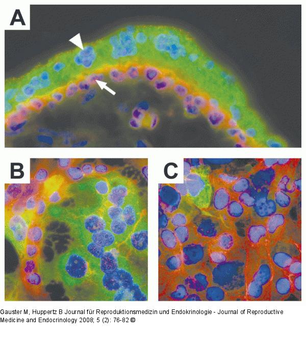
Abbildung 2a-c: Human placenta A) Immunofluorescent staining ofhuman first trimester placenta (week 8).Mononucleated cytotrophoblasts (arrow)show staining for E-cadherin in their apicaland lateral cell membranes (red). The over-lying multinucleated syncytiotrophoblast(arrowhead) lacks lateral cell walls and isnegative for E-cadherin. The syncytiotro-phoblast expresses hCG (green). B) and C)Immunofluorescent staining of forskolin andvehicle treated BeWo cells. Treatment ofBeWo cells for 48h with forskolin (20 μM)induced formation of multinucleated syn-cytia, visualized by degradation of E-cadherin (stained in red). hCG expression(green) was predominantly observed in syn-cytia of forskolin treated BeWo cells, butwas also rarely seen in mononucleatedBeWo cells (treated and untreated). |

Abbildung 2a-c: Human placenta
A) Immunofluorescent staining ofhuman first trimester placenta (week 8).Mononucleated cytotrophoblasts (arrow)show staining for E-cadherin in their apicaland lateral cell membranes (red). The over-lying multinucleated syncytiotrophoblast(arrowhead) lacks lateral cell walls and isnegative for E-cadherin. The syncytiotro-phoblast expresses hCG (green). B) and C)Immunofluorescent staining of forskolin andvehicle treated BeWo cells. Treatment ofBeWo cells for 48h with forskolin (20 μM)induced formation of multinucleated syn-cytia, visualized by degradation of E-cadherin (stained in red). hCG expression(green) was predominantly observed in syn-cytia of forskolin treated BeWo cells, butwas also rarely seen in mononucleatedBeWo cells (treated and untreated). |


