|
|
|---|
1. Schafhauser W;
Becher H;
Schaffhauser R;
Sachs M
Wird die Inzidenz von Lymphknotenmetastasen durch Sentinel-Lymphknoten-Dissektion beim Prostatakarzinom erhöht?
LN-Metastasen
Journal für Urologie und Urogynäkologie 2006; 13 (2) (Ausgabe für Österreich): 11-14
Journal für Urologie und Urogynäkologie 2006; 13 (2) (Ausgabe für Schweiz): 9-12
Journal für Urologie und Urogynäkologie 2006; 13 (2) (Ausgabe für Deutschland): 9-12
Detektionsrate von LN-Metastasen je nach Ausdehnung des Dissektionsgebietes (bei 32 SLN-positiven Pat. in 51 pelvinen Arealen) Keywords: Diagramm,
Metastase,
Urologie
|
|
|
2. Schafhauser W;
Becher H;
Schaffhauser R;
Sachs M
Wird die Inzidenz von Lymphknotenmetastasen durch Sentinel-Lymphknoten-Dissektion beim Prostatakarzinom erhöht?
Lymphknotenmetastasen - SLND
Journal für Urologie und Urogynäkologie 2006; 13 (2) (Ausgabe für Österreich): 11-14
Journal für Urologie und Urogynäkologie 2006; 13 (2) (Ausgabe für Schweiz): 9-12
Journal für Urologie und Urogynäkologie 2006; 13 (2) (Ausgabe für Deutschland): 9-12
Prozentsatz der durch SLND detektierten Lymphknotenmetastasen in Abhängigkeit vom Tumorstadium (n = 161 Patienten). Keywords: Diagramm,
Lymphom,
SLND,
Tumor,
Urologie
|
|
|
3. Schafhauser W;
Becher H;
Schaffhauser R;
Sachs M
Wird die Inzidenz von Lymphknotenmetastasen durch Sentinel-Lymphknoten-Dissektion beim Prostatakarzinom erhöht?
Pelvine Lymphszintigramme
Journal für Urologie und Urogynäkologie 2006; 13 (2) (Ausgabe für Österreich): 11-14
Journal für Urologie und Urogynäkologie 2006; 13 (2) (Ausgabe für Schweiz): 9-12
Journal für Urologie und Urogynäkologie 2006; 13 (2) (Ausgabe für Deutschland): 9-12
Kaplan-Meier-Analysen tumorunspezifisches im Vergleich mit Daten für pT4-Karzinome (aus [10]) und tumorspezifisches Überleben postoperativ. Keywords: Lymphom,
Szintigraphie,
Urologie
|
|
|
4. Kuntz-Hehner St;
Becher H;
Luederitz B;
Omran H;
Schlosser Th;
Tiemann K
Assessment of Myocardial Perfusion by Contrast Echocardiography - Ready for Clinical Practice?
Kontrastechokardiographie
Journal of Clinical and Basic Cardiology 2002; 5 (2): 145-148
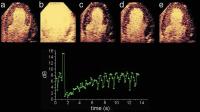
Negative bolus destruction reperfusion study using power pulse inversion imaging. First, continuous infusion of a contrast agent is adjusted to achieve full myocardial opacification using real-time imaging at very low emission power (a) (MI = 0.09). After destruction of the microbubbles by a short period of high power imaging (b) (MI = 1.3) myocardial contrast replenishment is recorded at low emission power (c–e) (MI = 0.09). A region of interest (ROI) is placed in the septum and repeated
measurements are made at the endsystolic frames. The graph shows the reperfusion curve for this ROI, fitting well to the monoexponential function. Keywords: Contrast agent,
echocardiography,
Echokardiographie,
Kontrastmittel,
reperfusion,
reperfusion
|
|
|
5. Kuntz-Hehner St;
Becher H;
Luederitz B;
Omran H;
Schlosser Th;
Tiemann K
Assessment of Myocardial Perfusion by Contrast Echocardiography - Ready for Clinical Practice?
Kontrastechokardiographie
Journal of Clinical and Basic Cardiology 2002; 5 (2): 145-148
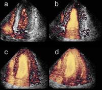
Different degrees of myocardial contrast using harmonic power Doppler imaging (four chamber view) during continuous infusion of Levovist (R): Arrival of the contrast agent in the right ventricle (a) and complete left ventricular opacification shortly after (b) using triggered imaging every heart cycle (1:1). Complete left ventricular myocardial opacification using higher trigger intervals every 3rd (c) and every 5th (d) cardiac cycle. Keywords: Contrast agent,
echocardiography,
Echokardiographie,
Four chamber view,
Kontrastmittel,
Levovist,
Levovist,
Schering GesmbH,
Schering GesmbH,
Vierkammerblick
|
|
|
6. Illien S;
Becher H;
Lüderitz B;
MacCarter D;
Omran H;
Rabahieh R;
Schimpf R;
Schmidt H;
Tiemann K;
von der Recke G
A Prospective Comparison of Harmonic Transthoracic and Transesophageal Echocardiography for Identifying Left Atrial Thrombi in Patients with Atrial Flutter and/or Fibrillation Prior to Cardioversion
Echokardiographie - Linksatriale Thromben
Journal of Clinical and Basic Cardiology 2002; 5 (1): 93-99
Transthoracic visualization of an LA appendage thrombus using a parasternal (A) and apical view (B). Arrows indicate thrombi. Keywords: Atrium,
Atrium,
echocardiography,
Echokardiographie,
Thrombus,
Thrombus
|
|
|
7. Illien S;
Becher H;
Lüderitz B;
MacCarter D;
Omran H;
Rabahieh R;
Schimpf R;
Schmidt H;
Tiemann K;
von der Recke G
A Prospective Comparison of Harmonic Transthoracic and Transesophageal Echocardiography for Identifying Left Atrial Thrombi in Patients with Atrial Flutter and/or Fibrillation Prior to Cardioversion
Echokardiographie - Linksatriale Thromben
Journal of Clinical and Basic Cardiology 2002; 5 (1): 93-99
Transthoracic recording of the velocity profile over the LA appendage in a patient with AF (A) and AFL (B). Keywords: Atrium,
Atrium,
echocardiography,
Echokardiographie,
Thrombus,
Thrombus
|
|
|
8. Illien S;
Becher H;
Lüderitz B;
MacCarter D;
Omran H;
Rabahieh R;
Schimpf R;
Schmidt H;
Tiemann K;
von der Recke G
A Prospective Comparison of Harmonic Transthoracic and Transesophageal Echocardiography for Identifying Left Atrial Thrombi in Patients with Atrial Flutter and/or Fibrillation Prior to Cardioversion
Echokardiographie - Linksatriale Thromben
Journal of Clinical and Basic Cardiology 2002; 5 (1): 93-99
Transthoracic imaging of the LA appendage using a parasternal view (A) and apical view (B). Arrow indicates LA appendage. Keywords: Atrium,
Atrium,
echocardiography,
Echokardiographie,
Thrombus,
Thrombus
|
|
|
| |

 Suche Medizinische Publikationen
 Suche Abbildungen und Grafiken
 Medizinprodukte - Labortechnik
 Antidotarium (Stand 2012)
 Phytokodex (Stand 2012)
 Fachbücher
 Patientenliteratur