| |
| Alle Journale wurden nach Chiariello L durchsucht |
Treffer 1 - 6 von 6 |
|
|
|---|
1. Gaspardone A;
Chiariello L;
Crea F;
De Matteis GM;
Gioffrè PA;
Polisca P;
Proietti I;
Tomai F;
Versaci F
Temporal Sequence and Spatial Distribution of Ischaemic Changes During Dipyridamole Stress Test - the Key Role of Microvascular Dysfunction
Different ischaemic events
Journal of Clinical and Basic Cardiology 2005; 8 (1-4): 47-53
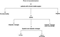
Flow chart showing the different ischaemic events in the study group during dipyridamole infusion after coronary artery revascularisation.
Numbers indicate the number of patients. ECG: electrocardiographic changes; ECHO: echocardiographic changes; LAD-dep: systolic changes in left anterior descending coronary artery-dependent territories; non-LAD-dep: systolic changes in left anterior descending coronary artery-independent territories; LAD-dep and non-LAD-dep: systolic changes in left anterior descending coronary artery-dependent and -independent territories. Keywords: ischaemia,
Ischämie,
revascularisation,
Revaskularisation,
Schema,
scheme
|
|
|
2. Gaspardone A;
Chiariello L;
Crea F;
De Matteis GM;
Gioffrè PA;
Polisca P;
Proietti I;
Tomai F;
Versaci F
Temporal Sequence and Spatial Distribution of Ischaemic Changes During Dipyridamole Stress Test - the Key Role of Microvascular Dysfunction
Different ischaemic events
Journal of Clinical and Basic Cardiology 2005; 8 (1-4): 47-53
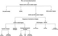
Flow chart showing the different ischaemic events in the study group during dipyridamole infusion before coronary artery revascularisation.
Numbers indicate the number of patients. ECG: electrocardiographic changes; ECHO: echocardiographic changes; LAD-dep:
systolic changes in left anterior descending coronary artery-dependent territories; non-LAD-dep: systolic changes in left anterior descending
coronary artery independent territories; LAD-dep and non-LAD-dep: systolic changes in left anterior descending coronary artery-dependent
and independent territories. Keywords: ischaemia,
Ischämie,
Schema,
scheme
|
|
|
3. Gaspardone A;
Chiariello L;
Crea F;
De Matteis GM;
Gioffrè PA;
Polisca P;
Proietti I;
Tomai F;
Versaci F
Temporal Sequence and Spatial Distribution of Ischaemic Changes During Dipyridamole Stress Test - the Key Role of Microvascular Dysfunction
Ischaemic events - Temporal sequence
Journal of Clinical and Basic Cardiology 2005; 8 (1-4): 47-53
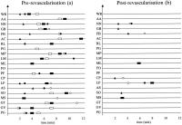
Schematic representation of the temporal sequence of ischaemic events during dipyridamole infusion in each patient before (panel
a) and after (panel b) coronary artery revascularisation. Black squares: angina pectoris; white squares: electrocardiographic ischaemic
changes; black circles: echocardiographic alterations in non-LAD-dependent myocardial segments; white circles: echocardiographic alterations
in LAD-dependent myocardial segments; black triangles: Doppler mitral flow abnormality. LAD: left anterior descending coronary artery Keywords: ischaemia,
Ischämie,
Schema,
scheme
|
|
|
4. Gaspardone A;
Chiariello L;
Crea F;
De Matteis GM;
Gioffrè PA;
Polisca P;
Proietti I;
Tomai F;
Versaci F
Temporal Sequence and Spatial Distribution of Ischaemic Changes During Dipyridamole Stress Test - the Key Role of Microvascular Dysfunction
Wall motion analysis
Journal of Clinical and Basic Cardiology 2005; 8 (1-4): 47-53
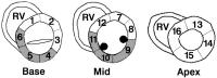
Sixteen-segment model for wall motion analysis. White segments were considered LAD-dependent myocardial segments (n = 10); shaded segments were considered non-LAD-dependent
myocardial segments (n = 6). 1: proximal anterior septum; 2: proximal anterior wall; 3: proximal lateral wall; 4: proximal posterior wall;
5: proximal inferior wall; 6: proximal inferior septum; 7: mid anterior wall; 8: mid lateral wall; 9: mid posterior wall; 10: mid inferior wall;
11: mid inferior septum; 12: mid anterior septum; 13: distal anterior wall; 14: distal lateral wall; 15: distal posterior wall; 16: distal anterior
septum. RV: right ventricle Keywords: LAD,
Schema,
scheme,
wall motion analysis
|
|
|
5. Gaspardone A;
Chiariello L;
Crea F;
Ferri C;
Gallo P;
Ghini AS;
Gioffrè PA;
Santucci A;
Tomai F;
Versaci F
The Enhanced Vasoreactivity of the Culprit Lesion in Unstable Angina is Associated with an Increased Local Release of Endothelin-1
Stenting - Endothelin-1
Journal of Clinical and Basic Cardiology 2002; 5 (1): 87-92
Endothelin-1 levels in blood sampled proximally and distally to the site of dilatation, before (Pre-stenting) and after (Poststenting) coronary artery stent implantation in patients with stable (shaded bars) and unstable (striped bars) angina. *: significantly different vs baseline (pre-stenting); +: significantly different unstable vs stable angina. Keywords: Concentration,
Diagramm,
endothelin-1,
endothelin-1,
instabile Angina pectoris,
Konzentration,
Stabile Angina pectoris,
stable angina pectoris,
Stent,
Stent,
unstable angina pectoris
|
|
|
6. Gaspardone A;
Chiariello L;
Crea F;
Ferri C;
Gallo P;
Ghini AS;
Gioffrè PA;
Santucci A;
Tomai F;
Versaci F
The Enhanced Vasoreactivity of the Culprit Lesion in Unstable Angina is Associated with an Increased Local Release of Endothelin-1
Stenose - Durchmesser
Journal of Clinical and Basic Cardiology 2002; 5 (1): 87-92
Plot of the luminal diameter of the coronary stenosis (A) and of the proximal (B), middle (C) and distal (D) angiographically normal-appearing coronary segments at baseline, during cold pressor test (CPT) and after intracoronary nitroglycerine administration (NTG). Black circles indicate patients with unstable angina and white circles indicate patients with stable angina. *: significantly different vs baseline; +: significantly different unstable vs stable angina. Keywords: Cold Pressure Test,
Cold Pressure Test,
culprit lesion,
Culprit-Läsion,
Diagramm,
Diameter,
Durchmesser,
Kältetest,
Lumen,
Lumen,
nitroglycerine,
Nitroglyzerin,
Stenose,
Stenosis
|
|
|
| |
|

 Suche Medizinische Publikationen
 Suche Abbildungen und Grafiken
 Medizinprodukte - Labortechnik
 Antidotarium (Stand 2012)
 Phytokodex (Stand 2012)
 Fachbücher
 Patientenliteratur