| |
| Alle Journale wurden nach Erbel R durchsucht |
Treffer 1 - 11 von 11 |
|
|
|---|
1. Bartel T;
Caspari G;
Erbel R;
Mueller S
Intracardiac Echocardiography - Technology and Clinical Role
Intrakardiale Echokardiographie
Journal of Clinical and Basic Cardiology 2002; 5 (2): 133-137
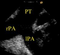
Left and right pulmonary arteries viewed with the
AcuNav (TM)-catheter in the pulmonary trunk. lPA = left pulmonary artery; rPA = right pulmonary artery; PT = pulmonary trunk Keywords: Arteria pulmonalis,
Arteria pulmonalis,
Catheter,
echocardiography,
Echokardiographie,
Katheter,
Truncus pulmonalis,
Truncus pulmonalis
|
|
|
2. Bartel T;
Caspari G;
Erbel R;
Mueller S
Intracardiac Echocardiography - Technology and Clinical Role
Intrakardiale Echokardiographie
Journal of Clinical and Basic Cardiology 2002; 5 (2): 133-137
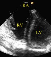
The right ventricle viewed with the AcuNav (TM)-catheter pressed against the roof of the right atrium. LV = left ventricle; RA = right atrium; RV = right ventricle Keywords: Atrium,
Atrium,
Catheter,
echocardiography,
Echokardiographie,
Katheter,
ventricle,
Ventrikel
|
|
|
3. Bartel T;
Caspari G;
Erbel R;
Mueller S
Intracardiac Echocardiography - Technology and Clinical Role
Intrakardiale Echokardiographie
Journal of Clinical and Basic Cardiology 2002; 5 (2): 133-137
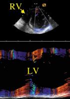
The AcuNav (TM)-catheter is now pressed against the lateral RV wall avoiding its motion within the right ventricular cavity. LV = left ventricle; RV = right ventricle Keywords: Catheter,
echocardiography,
Echokardiographie,
Katheter,
ventricle,
Ventrikel
|
|
|
4. Bartel T;
Caspari G;
Erbel R;
Mueller S
Intracardiac Echocardiography - Technology and Clinical Role
Intrakardiale Echokardiographie
Journal of Clinical and Basic Cardiology 2002; 5 (2): 133-137
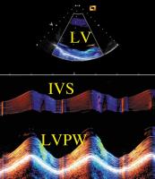
Rhythmic motion of the AcuNav (TM)-catheter within the right ventricle causes pseudoparadoxical motion of the interatrial septum. IVS = interventricular septum; LV = left ventricle; LVPW =
left ventricular posterior wall Keywords: Catheter,
echocardiography,
Echokardiographie,
Katheter,
Septum,
Septum,
ventricle,
Ventrikel
|
|
|
5. Bartel T;
Caspari G;
Erbel R;
Mueller S
Intracardiac Echocardiography - Technology and Clinical Role
Intrakardiale Echokardiographie
Journal of Clinical and Basic Cardiology 2002; 5 (2): 133-137
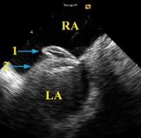
Device closure of a patent foramen ovale guided by
intracardiac echocardiography with the AcuNav )(TM)-catheter placed in the right atrium and retroflexed into a short-axis orientation. Typical attitude before the Amplatzer (TM)-PFO occluder is removed from the cable. LA = left atrium; RA = right atrium; 1, right umbrella of Amplatzer (TM)-PFO occluder; 2, left umbrella of Amplatzer (TM)-PFO occluder Keywords: Amplatzer-PFO-Occluder,
Amplatzer-PFO-Occluder,
Atrium,
Atrium,
Catheter,
echocardiography,
Echokardiographie,
Foramen ovale,
Foramen ovale,
Katheter
|
|
|
6. Bartel T;
Caspari G;
Erbel R;
Mueller S
Intracardiac Echocardiography - Technology and Clinical Role
Intrakardiale Echokardiographie
Journal of Clinical and Basic Cardiology 2002; 5 (2): 133-137
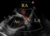
Color-Doppler image of a left-to-right shunt due to a small ASD. Ao = aorta; LA = left atrium; RA = right atrium Keywords: Aorta,
Aorta,
Atrium,
Atrium,
Colordoppler,
echocardiography,
Echokardiographie,
Farbdoppler,
Shunt,
Shunt
|
|
|
7. Bartel T;
Caspari G;
Erbel R;
Mueller S
Intracardiac Echocardiography - Technology and Clinical Role
Intrakardialer Ultraschall
Journal of Clinical and Basic Cardiology 2002; 5 (2): 133-137
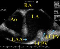
Left atrium, left atrial appendage, and left pulmonary veins demonstrated with the AcuNav(TM)-catheter being placed in the right atrium and lined up with the interatrial septum. Ao = aorta; LAA = left atrial appendage; LA = left atrium; LLPV = left lower pulmonary vein; LUPV = left upper pulmonary vein; RA = right atrium Keywords: Arteria pulmonalis,
Arteria pulmonalis,
Atrium,
Atrium,
Catheter,
echocardiography,
Echokardiographie,
Katheter
|
|
|
|
|
|
9. Bartel T;
Caspari G;
Erbel R;
Mueller S
Intracardiac Echocardiography - Technology and Clinical Role
Intrakardiale Echokardiographie
Journal of Clinical and Basic Cardiology 2002; 5 (2): 133-137
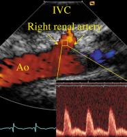
Right renal artery viewed from the inferior vena cava including color Doppler flow and spectral Doppler analysis. Ao = aorta; IVC = inferior vena cava Keywords: Aorta abdominalis,
Aorta abdominalis,
Arteria renalis,
Arteria renalis,
Colordoppler,
echocardiography,
Echokardiographie,
Farbdoppler,
Vena cava inferior,
Vena cava inferior
|
|
|
10. Wenzel RR;
Baumgart D;
Bruck H;
Erbel R;
Heemann U;
Mitchell A;
Philipp Th;
Schaefers RF
Interaction of the Sympathetic Nervous System with other Pressor Systems in Antihypertensive Therapy
Moxonidin - Herzfrequenz
Journal of Clinical and Basic Cardiology 2001; 4 (3): 185-192
Effect of the central sympathicolytic moxonidine on heart rate (Details see text). Modified from reference [127] Keywords: Diagramm,
heart rate,
Herzfrequenz,
Moxonidin,
moxonidine
|
|
|
11. Wenzel RR;
Baumgart D;
Bruck H;
Erbel R;
Heemann U;
Mitchell A;
Philipp Th;
Schaefers RF
Interaction of the Sympathetic Nervous System with other Pressor Systems in Antihypertensive Therapy
Moxonidin - SNS-Überaktivität
Journal of Clinical and Basic Cardiology 2001; 4 (3): 185-192
Effect of the central sympathicolytic moxonidine on central SNS activity (muscle sympathetic nerve activity) as assessed by microneurography. Modified from reference [127] Keywords: Activity,
Aktivität,
Diagramm,
Microneurography,
Mikroneurographie,
Moxonidin,
moxonidine,
sympathetic nervous system,
Sympathikus,
sympathisches Nervensystem
|
|
|
| |
|

 Suche Medizinische Publikationen
 Suche Abbildungen und Grafiken
 Medizinprodukte - Labortechnik
 Antidotarium (Stand 2012)
 Phytokodex (Stand 2012)
 Fachbücher
 Patientenliteratur