| |
| Alle Journale wurden nach Huppertz B durchsucht |
Treffer 1 - 7 von 7 |
|
|
|---|
1. Gauster M;
Huppertz B
Fusion of Cytothrophoblast with Syncytiotrophoblast in the Human Placenta: Factors Involved in Syncytialization
Trophoblast fusion
Journal für Reproduktionsmedizin und Endokrinologie - Journal of Reproductive Medicine and Endocrinology 2008; 5 (2): 76-82
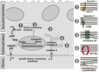
Illustration of factors involved in trophoblast fusion. Environmentally derived growthfactors, hormones and cytokines bind to their cognate receptors at the plasma membrane ofcytotrophoblasts. Activation of either protein kinase A (PKA) or MAP kinases ERK1/2 and p38leads to increased protein expression of the transcription factor GCMa, which in turn drivestranscription of fusogenic genes. Several structural and membrane proteins were suggested topromote trophoblast fusion, including syncytin 1 and its receptor ASCT2 (1), CD98 and its receptorgalectin 3 (2) as well as connexin 43 (3). Beside activation of PKA or MAP kinase pathways,cytokines induce conversion of pro-caspase 8 into active caspase 8. Once activated, caspase 8can mediate inactivation of “flippasesâ€� and/or activation of "floppases" to trigger phosphatidyl-serine externalisation (PS flip) (4). Additionally, caspase 8 triggers remodelling of the sub-mem-branous cytoskeleton by degrading structural proteins such as α-fodrin (5). Keywords: Throphoblast,
trophoblast fusion
|
|
|
2. Gauster M;
Huppertz B
Fusion of Cytothrophoblast with Syncytiotrophoblast in the Human Placenta: Factors Involved in Syncytialization
Human placenta
Journal für Reproduktionsmedizin und Endokrinologie - Journal of Reproductive Medicine and Endocrinology 2008; 5 (2): 76-82
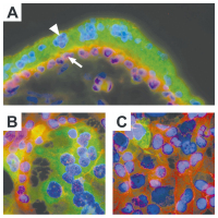
A) Immunofluorescent staining ofhuman first trimester placenta (week 8).Mononucleated cytotrophoblasts (arrow)show staining for E-cadherin in their apicaland lateral cell membranes (red). The over-lying multinucleated syncytiotrophoblast(arrowhead) lacks lateral cell walls and isnegative for E-cadherin. The syncytiotro-phoblast expresses hCG (green). B) and C)Immunofluorescent staining of forskolin andvehicle treated BeWo cells. Treatment ofBeWo cells for 48h with forskolin (20 μM)induced formation of multinucleated syn-cytia, visualized by degradation of E-cadherin (stained in red). hCG expression(green) was predominantly observed in syn-cytia of forskolin treated BeWo cells, butwas also rarely seen in mononucleatedBeWo cells (treated and untreated). Keywords: placenta,
Plazenta
|
|
|
3. Gauster M;
Huppertz B
Fusion of Cytothrophoblast with Syncytiotrophoblast in the Human Placenta: Factors Involved in Syncytialization
Trophoblast
Journal für Reproduktionsmedizin und Endokrinologie - Journal of Reproductive Medicine and Endocrinology 2008; 5 (2): 76-82
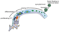
Schematic representation of trophoblast differentiation within the villous trophoblast.Derived from trophoblast progenitor cells daughter cells start to differentiate and finally fusewith the overlying syncytiotrophoblast. Nuclei that have become an integrative part of the syn-cytiotrophoblast change their morphology and display chromatin condensation. Finally lateapoptotic syncytial nuclei are packed into syncytial knots and are released into the maternalblood stream. The arrows follow the route of cell types and nuclear changes during trophoblastdifferentiation. Keywords: Schema,
scheme,
Trohoblast,
trophoblast
|
|
|
4. Huppertz B
Placental Villous Trophoblast: the Altered Balance Between Proliferation and Apoptosis Triggers Pre-eclampsia
Term placentas
Journal für Reproduktionsmedizin und Endokrinologie - Journal of Reproductive Medicine and Endocrinology 2006; 3 (2): 103-108
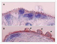
Representative images of chorionic villi from term placentas derived from a normal pregnancy (A) and a pregnancy complicated by late-onset pre-eclampsia (B). In both images staining was performed using a primary antibody against placental-protein 13 (PP13), which is present in the apical membrane of the syncytiotrophoblast. In (A) the normal villous brush border can be seen, which is stained for PP13. In (B) the alterations of the brush border membrane are obvious. Protrusions
on the cellular level can be seen (arrows) that may be released into the maternal circulation. Magnification ×900. Keywords: Plazenta,
pre-eclampsia,
pregnancy,
Präeklampsie,
Schwangerschaft,
term placentas
|
|
|
5. Huppertz B
Placental Villous Trophoblast: the Altered Balance Between Proliferation and Apoptosis Triggers Pre-eclampsia
Trophoblast turnover
Journal für Reproduktionsmedizin und Endokrinologie - Journal of Reproductive Medicine and Endocrinology 2006; 3 (2): 103-108
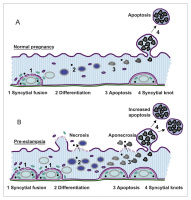
Schematic representation of trophoblast turnover. (A) Normal pregnancy: The final event of cytotrophoblast differentiation, syncytial fusion, results in incorporation of fresh organelles and other cellular
material into the syncytium (1). Within the multinucleated syncytiotrophoblast
differentiation (2) and subsequently late apoptosis (3) take place. Finally, the late apoptotic material is packed into protrusions of the apical plasma membrane, syncytial knots. These knots are released into the maternal circulation as tightly sealed corpuscles (4). (B) Pre-eclampsia: Enhanced proliferation and syncytial fusion (1) may overwhelm the
capacity of the syncytiotrophoblast in terms of differentiation (2) and
apoptotic release (3). This may result in a necrotic breakdown of specific
sites of the syncytiotrophoblast. If at these sites apoptosis has not yet started, pure necrosis can be observed; if however apoptosis has already lead to first cleavage of proteins, apoptotic material will be necrotically released (aponecrosis). At the same time the syncytiotrophoblast tries to counter balance for the increased input by increasing the release of apoptotic syncytial knots (4). Modified from [15]. Keywords: pregnancy,
Schema,
scheme,
Schwangerschaft,
trophoblast
|
|
|
6. Huppertz B
Das Epithel der Plazentazotten: Veränderungen des Trophoblasten bei Präeklampsie
Trophoblast
Journal für Reproduktionsmedizin und Endokrinologie - Journal of Reproductive Medicine and Endocrinology 2005; 2 (2): 84-89
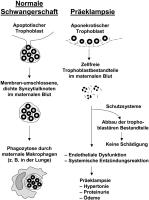
Die Qualität der Abgabe trophoblastären Materials. Links ist die normale apoptotische Abgabe gezeigt. Alte Kerne und anderes gealtertes Material werden in Synzytialknoten gesammelt und als Membran-
umschlossene Gebilde nach außen in die maternale Zirkulation abgegeben. Ohne daß intrazelluläre Bestandteile nach außen gelangen, werden diese Strukturen von Makrophagen phagozytiert und somit aus der maternalen Blutbahn entfernt. Rechts ist die pathologische Freisetzung trophoblastären Materials bei Präeklampsie gezeigt. Hier wird apoptotisches Material nekrotisch freigesetzt (Aponekrose) und gelangt somit zellfrei in die maternale Blutbahn. Solange maternale Schutzsysteme dieses Material abfangen können, bleibt die Mutter ohne Schädigung. Bei einer Überschreitung der Kapazitäten der Schutzmechanismen der Mutter kommt es zu Schädigungen des Endothels mit nachfolgenden Symptomen bis hin zur Ausprägung der Präeklampsie. Mod. nach [1]. Keywords: Embryologie,
Plazenta,
Präeklampsie,
Reproduktionsmedizin,
Schema,
Trophoblasten
|
|
|
7. Huppertz B
Das Epithel der Plazentazotten: Veränderungen des Trophoblasten bei Präeklampsie
Villösen Trophoblasten - Differenzierung
Journal für Reproduktionsmedizin und Endokrinologie - Journal of Reproductive Medicine and Endocrinology 2005; 2 (2): 84-89
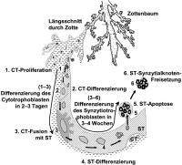
Verlauf der Differenzierung des villösen Trophoblasten. Der Zottenbaum oben in der Abbildung repräsentiert einen Teil des gesamten Zottengewebes der Plazenta. Ausgehend davon findet sich von links nach unten ziehend ein Längsschnitt durch eine Zotte, bei der im unteren Bildteil vor allem das Zottenepithel, der villöse Trophoblast, hervorgehoben ist. In dieses Epithel sind die einzelnen Schritte der Differenzierung und des Umsatzes durch die Zahlen 1–6 symbolisiert und durch die schwarzen Pfeile der Verlauf gekennzeichnet. 1: Die Stammzellen im Verband des Zytotrophoblasten (CT) proliferieren und generieren Tochterzellen. 2: Diese mononukleären Tochterzellen verlassen den Zellzyklus und differenzieren, bis sie schließlich als letzten Schritt ihrer Differenzierung (3) mit dem Synzytiotrophoblasten (ST) fusionieren. 4: Innerhalb des Synzytiotrophoblasten kommt es zu weiteren Differenzierungsvorgängen, um den hohen Funktionsstatus dieses Gewebes aufrecht zu erhalten. 5: Letztendlich altern die Kerne des Synzytiotrophoblasten und werden in Synzytialknoten gesammelt. 6: Diese Synzytialknoten werden vom Synzytiotrophoblasten abgeschnürt und in die maternale Zirkulation abgegeben. Dort werden sie überwiegend in der Lunge der Mutter phagozytiert. Mod. nach [1]. Keywords: Embryologie,
Plazenta,
Reproduktionsmedizin,
Schema,
Trophoblasten
|
|
|
| |
|

 Suche Medizinische Publikationen
 Suche Abbildungen und Grafiken
 Medizinprodukte - Labortechnik
 Antidotarium (Stand 2012)
 Phytokodex (Stand 2012)
 Fachbücher
 Patientenliteratur