| |
| Alle Journale wurden nach Khan IA durchsucht |
Treffer 1 - 8 von 8 |
|
|
|---|
1. Gowda RM;
Caccavo ND;
Cosme-Thormann BF;
Khan IA
Single Coronary Artery: Anomalous Origin of Right Coronary Artery from Left Anterior Descending Artery
Coronary angiography
Journal of Clinical and Basic Cardiology 2005; 8 (1-4): 73
Coronary angiography shows origin of right coronary artery from left anterior descending coronary artery. Keywords: coronary angiography,
Koronarangiographie
|
|
|
2. Shaikh N;
Daya SK;
Gowda RM;
Khan IA;
Kundu S
Venacavo-Atrial Renal Tumor
Transesophageal Echocardiography
Journal of Clinical and Basic Cardiology 2004; 7 (1-4): 28-29
Transesophageal echocardiography in horizontal
view: A large mass is seen in the right atrium. Keywords: right atrium,
transesophageal echocardiography
|
|
|
3. Shaikh N;
Daya SK;
Gowda RM;
Khan IA;
Kundu S
Venacavo-Atrial Renal Tumor
Transthoracic echocardiography
Journal of Clinical and Basic Cardiology 2004; 7 (1-4): 28-29
Transthoracic echocardiogram in 4-chamber view demonstrates a large mass in the right atrium. Keywords: right atrium,
transthoracic echocardiography
|
|
|
4. Raufi A;
Khan IA;
Nair VM;
Rahmatullah SI;
Rodriguez C;
Sacchi TJ;
Sahni G;
Vasavada BC
Rupture of a Surgically Repaired Sinus of Valsalva Aneurysm
Sinus-Valsalvae-Aneurysma
Journal of Clinical and Basic Cardiology 2002; 5 (2): 199-200
Aortogram demonstrates a communication between aorta and right atrium (RA) Keywords: Aneurysma,
Aneurysma,
Aorta,
Aorta,
Aortogramm,
Aortogramm,
Atrium,
Atrium,
Shunt,
Shunt,
Sinus valsalvae,
Sinus valsalvae
|
|
|
5. Raufi A;
Khan IA;
Nair VM;
Rahmatullah SI;
Rodriguez C;
Sacchi TJ;
Sahni G;
Vasavada BC
Rupture of a Surgically Repaired Sinus of Valsalva Aneurysm
Sinus-Valsalvae-Aneurysma
Journal of Clinical and Basic Cardiology 2002; 5 (2): 199-200
Transoesophageal echocardiography demonstrates dilated right atrium (RA) and right ventricle (RV) and a high velocity shunt from aorta to right atrium. LA = left atrium, LV = left ventricle Keywords: Aneurysma,
Aneurysma,
Aorta,
Aorta,
Atrium,
Atrium,
echocardiography,
Echokardiographie,
Ruptur,
rupture,
Shunt,
Shunt,
Sinus valsalvae,
Sinus valsalvae,
TEE,
TEE,
ventricle,
Ventrikel
|
|
|
6. Siddiqui MA;
Khan IA
Transgastric and Transoesophageal Echocardiographic Views of Right Atrial Appendage
Rechtsatrialer Appendix
Journal of Clinical and Basic Cardiology 2001; 4 (4): 301
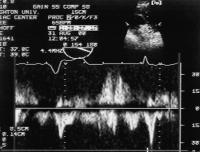
The pulsed Doppler evaluation of right atr ial appendage through 154 degree transgastric view. The pulsed Doppler sample volume is shown in right upper corner of the figure. Keywords: Appendix,
Atrium,
echocardiography,
Echokardiographie,
right atrial appendage
|
|
|
7. Siddiqui MA;
Khan IA
Transgastric and Transoesophageal Echocardiographic Views of Right Atrial Appendage
Rechtsatrialer Appendix
Journal of Clinical and Basic Cardiology 2001; 4 (4): 301
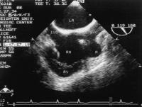
Transoesophageal echocardiographic view of both atr ia in longitudinal plane of 119 degree. The right atrial appendage (A) is visualized in its horizontal plane. IVC = inferior vena cava, LA = left atrium, RA = right atrium, RV = right ventricle, SVC = superior vena cava, TV = tricuspid valve, W = pacemaker wire Keywords: Appendix,
Atrium,
echocardiography,
Echokardiographie,
right atrial appendage
|
|
|
8. Siddiqui MA;
Khan IA
Transgastric and Transoesophageal Echocardiographic Views of Right Atrial Appendage
Rechtsatrialer Appendix
Journal of Clinical and Basic Cardiology 2001; 4 (4): 301
Transgastric echocardiographic view of right-sided card iac chambers in tangential plane of 153 degree. The right atrial appendage (A) is visualized in its longitudinal plane. RA = right atrium, RV = right ventricle, TV = tricuspid valve Keywords: Appendix,
Atrium,
echocardiography,
Echokardiographie,
right atrial appendage
|
|
|
| |
|

 Suche Medizinische Publikationen
 Suche Abbildungen und Grafiken
 Medizinprodukte - Labortechnik
 Antidotarium (Stand 2012)
 Phytokodex (Stand 2012)
 Fachbücher
 Patientenliteratur