|
|
|---|
|
|
|
|
|
|
3. Metzler B
Behandlung der myokardialen Dysfunktion nach kardiopulmonaler Reanimation // Treatment of Post-Cardiac Arrest Syndrome
Myokardiale Dysfunktion - Behandlung
Journal für Kardiologie - Austrian Journal of Cardiology 2015; 22 (5-6): 120-122
Vorgeschlagener Algorithmus zur Diagnose und Behandlung der myokardialen Dysfunktion nach kardiopulmonaler Reanimation. Keywords: kardiopulmonale Reanimation,
myokardiale Dysfunktion,
Schema
|
|
|
4. Mayr A;
Mueller L;
Kaufmann M;
Metzler B
Case Report: “High Take-Off� of the Left and Right Coronary Artery
Coronary artery
Journal für Kardiologie - Austrian Journal of Cardiology 2013; 20 (7-8): 216
Sagittal CT image showing the origin of the RCA (black arrowhead) from the anterior-superior aspect of the ascending aorta, 4.7 cm above the sinotubular junction. (B): Coronal images showing the origin of the LCA (black arrow) originated
from the left-lateral surface of the dilated tubular ascending aorta, 3.9 cm above its usual location. (C): Right lateral view of a Volume Rendered (VR) image showing the high take-off of the RCA (black arrowhead) and the LCA (white arrowhead). (D): Intraoperative situs showing the unusal origin of the RCA (black arrowhead). Keywords: angiography,
Aorta,
coronary artery,
CT,
Kardiologie,
photo
|
|
|
5. Mayr A;
Alber H;
Klug G;
Pachinger O;
Metzler B
Case Report: Hidden Dissection of the Left Descending Coronary Artery Mimicking Tako-Tsubo Cardiomyopathy
STEMI
Journal für Kardiologie - Austrian Journal of Cardiology 2012; 19 (9-10): 289-290
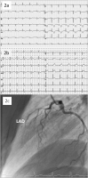
a): ECG one day after the first coronary angiography showing subacute
anterior STEMI; (b): ECG three days after the first coronary angiography showing
again significant anterior ST segment elevations; the patient once again developed
massive retrosternal chest pain; (c): Second coronary angiography revealed complete
occlusion of the proximal LAD due to spontaneous dissection. Keywords: Angiographie,
coronary angiography,
ECG,
EKG,
Kardiologie,
STEMI
|
|
|
6. Mayr A;
Alber H;
Klug G;
Pachinger O;
Metzler B
Case Report: Hidden Dissection of the Left Descending Coronary Artery Mimicking Tako-Tsubo Cardiomyopathy
STEMI
Journal für Kardiologie - Austrian Journal of Cardiology 2012; 19 (9-10): 289-290
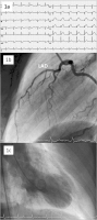
1. (a): Initial ECG showing anterior STEMI; (b): First coronary angiography
showing no obstruction/stenosis of the left descending coronary artery (LAD). Right
coronary artery and circumflex artery also showed no abnormalities; (c): Global left
ventricular function was reduced moderately with anterolateral, apical as well as
inferior hypokinesis. Keywords: angiography,
coronary angiography,
ECG,
EKG,
Kardiologie,
STEMI
|
|
|
7. Hörmann P;
Wanitschek M;
Metzler B;
Alber H
Milde therapeutische Hypotherapie beim Myokardinfarkt
Reperfusions-Hypothermie
Journal für Kardiologie - Austrian Journal of Cardiology 2012; 19 (9-10): 274-276
Auswirkungen von Prä- und Post-Reperfusions-Hypothermie auf die Infarktgröße im Schweinemodell. Mod. nach [21]. Keywords: Diagramm,
Kardiologie,
Reperfusions-Hypothermie
|
|
|
8. Metzler B
Definition des Hochrisikopatienten und therapeutische Optionen beim Patienten mit ST-Hebungsmyokardinfarkt
Kammerflimmern
Journal für Kardiologie - Austrian Journal of Cardiology 2010; 17 (7-8): 263-268
Algorithmus bei Kammerflimmern im Rahmen eines akuten Herzinfarktes:
Hierfür wird in vielen Zentren eine therapeutische Hypothermie gleichzeitig bzw.
unmittelbar nach interventioneller Verschlusseröffnung des infarktbezogenen Koronargefäßes
begonnen, obwohl dies derzeit noch nicht durch randomisierte Studien
geklärt ist bzw. durch Guidelines der internationalen Fachgesellschaften empfohlen
wird. Keywords: Kammerflimmern,
Kardiologie,
Schema
|
|
|
9. Klug G;
Trieb T;
Schocke M;
Nocker M;
Skalla E;
Nowosielski M;
Pedarnig K;
Mayr A;
Haubner B;
Pachinger O;
Metzler B
Morphologische und funktionelle Magnetresonanztomographie-Untersuchungen nach interventioneller Revaskularisierung bei akutem Myokardinfarkt
Infarzierte Segmente
Journal für Kardiologie - Austrian Journal of Cardiology 2008; 15 (3-4): 73-76
Balkendiagramm der regionalen Funktion infarzierter Segmente. Es zeigt sich kein signifikanter Unterschied zwischen den Gruppen bei der ersten Untersuchung (Baseline). Nach 3 Monaten verbesserten sich nur die Gruppen mit einer Ischämiezeit < 6 h bezüglich der regionalen Kontraktilität signifikant (*: p < 0,05;
+: p < 0,001; gepaarter Wilcoxon rank-Test). Der Betrag der Erholung der regionalen Funktion (deltaSWT) war in der Gruppe, welche innerhalb von 3 Stunden reperfundiert wurde, größer als in den Gruppen mit längeren Ischämiezeiten (#: p < 0,02; Kruskal-Wallis-Test). Daraus resultierte eine signifikant bessere Kontraktilität dieser
Segmente nach 4 Monaten (§: p < 0,01; Kruskal-Wallis-Test). Syst.: systolisch; EdWd:
Enddiastolische Wanddicke; n. s.: nicht signifikant. Keywords: Diagramm,
Infarziertes Segment,
Kardiologie
|
|
|
10. Klug G;
Trieb T;
Schocke M;
Nocker M;
Skalla E;
Nowosielski M;
Pedarnig K;
Mayr A;
Haubner B;
Pachinger O;
Metzler B
Morphologische und funktionelle Magnetresonanztomographie-Untersuchungen nach interventioneller Revaskularisierung bei akutem Myokardinfarkt
MRT-Datenauswertung
Journal für Kardiologie - Austrian Journal of Cardiology 2008; 15 (3-4): 73-76
MRT-Datenauswertung. Infarzierte Segmente wurden anhand der späten Kontrastmittelaufnahme identifiziert (a). Der Infarkt in den Segmenten 3, 4, 9, 10 und 15 führt zu einer deutlichen Beeinträchtigung der regionalen systolischen Wanddickenzunahme (b). Rote bis grüne Areale zeigen eine vorhandene Kontraktionsfähigkeit
des Myokards, während blaue bis schwarze Bereiche hypo- bis akinetisch sind. Die Einteilung der myokardialen Segmente erfolgte nach der AHA-Klassifikation (c). Die rote Linie verdeutlicht den vorderen Ansatzpunkt des rechten an dem linken
Ventrikel. Dieser Punkt dient als Referenzpunkt für die Segmenteinteilung. Keywords: Datenauswertung,
Kardiologie,
Kontrastmittel,
MRT
|
|
|
11. Conci E;
Pachinger O;
Metzler B
Mouse Models for Myocardial Ischaemia/Reperfusion
TTC-stained heart
Journal für Kardiologie - Austrian Journal of Cardiology 2006; 13 (7-8): 239-244
Cross sections of the TTC-stained heart. a) Areas at risk are delineated by Evans blue dye in a representative heart with occlusion of the left descending coronary artery. All areas that stain blue were therefore not at risk. b) Areas that appear pale white are infarcted. Areas that remain red were not infarcted but were within
the area at risk. Transverse sections are from the apex to the base (from left to right side). Keywords: Evans,
Kardiologie,
TTC
|
|
|
12. Conci E;
Pachinger O;
Metzler B
Mouse Models for Myocardial Ischaemia/Reperfusion
Mouse model
Journal für Kardiologie - Austrian Journal of Cardiology 2006; 13 (7-8): 239-244
Open mouse chest with occluded left descending coronary artery. Photograph of open chest
mouse with occluded left descending
coronary artery by the help of a PE-10 plastic tubing. The LAD ligation was done with an 8-0 silk and was evident by discoloration of the
left ventricle. Arrows indicate the
area at risk. Keywords: Foto,
Kardiologie,
linke Koronararterie,
Mausmodell
|
|
|
13. Conci E;
Pachinger O;
Metzler B
Mouse Models for Myocardial Ischaemia/Reperfusion
Adhesion Molecules
Journal für Kardiologie - Austrian Journal of Cardiology 2006; 13 (7-8): 239-244
Scheme of involvement of the adhesion molecules in ischaemia/reperfusion. Under normal conditions, there are virtually no interactions between the endothelium and neutrophils. After coronary ischaemia/reperfusion, the blood vessel’s ability
to form NO is severely reduced. With decreased supplies of NO, the coronary vessel
may constrict. Additional adhesion molecules were expressed on the endothelium and polymorphonuclear leukocytes (PMNs) begin with rolling as the first step in PMN
sequestration. As the PMN slows, tighter interactions between CD18 (on PMN) and
ICAM-1 (on endothelium) cause the second phase of the PMN-EC interaction called sticking. The final step, the diapedesis, involves the coordination of many factors. Keywords: Kardiologie,
myokardiale Ischämie,
Perfusion,
Schema
|
|
|
14. Metzler B;
Bilgeri R;
Pachinger O
Fulminant course of hypertension caused by phaeochromocytoma
Phäochromozytom - Blutdruck
Journal of Clinical and Basic Cardiology 2000; 3 (1): 67-68
Thirteen-hour systolic and diastolic blood pressure monitoring graph of an antihypertensive treated patient with an unknown Phaeochromocytoma. The x axis indicates the time after admission to hospital (h); the y-axis the blood pressure in mmHg. Keywords: blood pressure,
Blutdruck,
Diagramm,
hypertension,
Hypertonie,
phaeochromocytoma,
Phäochromozytom
|
|
|
| |

 Suche Medizinische Publikationen
 Suche Abbildungen und Grafiken
 Medizinprodukte - Labortechnik
 Antidotarium (Stand 2012)
 Phytokodex (Stand 2012)
 Fachbücher
 Patientenliteratur