Kopp AF, Claussen CD, Heuschmid M, Kuettner A, Schroeder S New Developments in Cardiac Imaging: The Role of MDCT Journal of Clinical and Basic Cardiology 2001; 4 (4): 253-260 PDF Summary Overview
| ||||||||||||
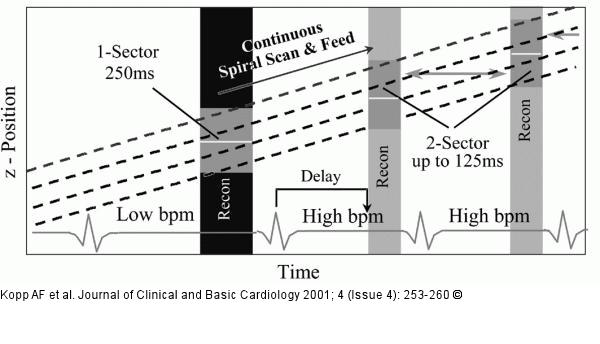
Figure/Graphic 2: Computertomographie - Bildrekonstruktion Retrospectively ECG-gated 4-slice spiral reconstruction with 1-sector reconstruction (black bar) for heart rates < 70 bpm and 2-sector reconstruction (2 grey bars are used for one reconstruction) for heart rates >= 70 bpm. The dashed lines indicate how the four detector rows travel along z-axis at a fixed speed (pitch). Using the adaptive approach gap-less volume reconstruction is possible with pitch 1.5 for all heart rates > 40 bpm. |

Figure/Graphic 2: Computertomographie - Bildrekonstruktion
Retrospectively ECG-gated 4-slice spiral reconstruction with 1-sector reconstruction (black bar) for heart rates < 70 bpm and 2-sector reconstruction (2 grey bars are used for one reconstruction) for heart rates >= 70 bpm. The dashed lines indicate how the four detector rows travel along z-axis at a fixed speed (pitch). Using the adaptive approach gap-less volume reconstruction is possible with pitch 1.5 for all heart rates > 40 bpm. |


