Kopp AF, Claussen CD, Heuschmid M, Kuettner A, Schroeder S New Developments in Cardiac Imaging: The Role of MDCT Journal of Clinical and Basic Cardiology 2001; 4 (4): 253-260 PDF Summary Overview
| ||||||||||||
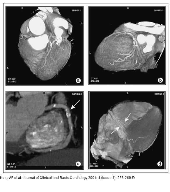
Figure/Graphic 3a-d: MDCT-Computertomographie MDCT angiography (collimation 4 x 1 mm, pitch 1.5, 120 cc Imeron(R) 400). (a) Anterior view of left coronary artery with LAD in volume rendering technique. (b) Lateral view of left coronary artery with LAD and circumflex branch. (c) Maximum intensity projection of right coronary artery with calcified plaques (arrow). (d) Diaphragmatic surface with posterolateral and interventricular branches of RCA (arrows). |

Figure/Graphic 3a-d: MDCT-Computertomographie
MDCT angiography (collimation 4 x 1 mm, pitch 1.5, 120 cc Imeron(R) 400). (a) Anterior view of left coronary artery with LAD in volume rendering technique. (b) Lateral view of left coronary artery with LAD and circumflex branch. (c) Maximum intensity projection of right coronary artery with calcified plaques (arrow). (d) Diaphragmatic surface with posterolateral and interventricular branches of RCA (arrows). |


