Kopp AF, Claussen CD, Heuschmid M, Kuettner A, Schroeder S New Developments in Cardiac Imaging: The Role of MDCT Journal of Clinical and Basic Cardiology 2001; 4 (4): 253-260 PDF Summary Overview
| ||||||||||||
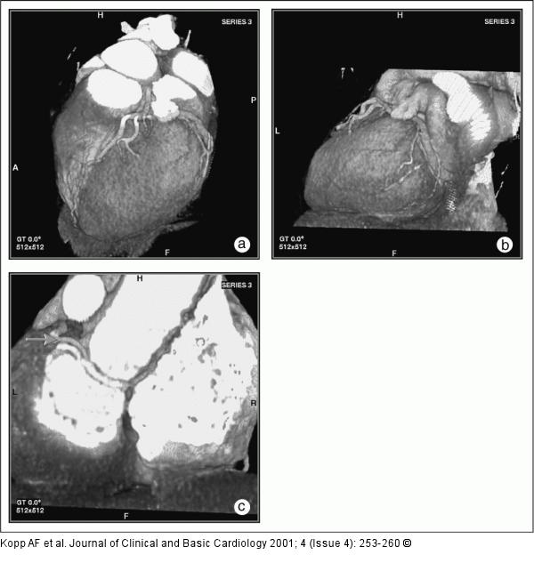
Figure/Graphic 4a-c: MCDT - Computertomographie Male 72-year old patient with known coronary anomaly (RCX originates from right coronary sinus) and absent CAD. Patient presented with recurrent chestpain. Stress-ECG up to 250 W showed no signs of ischaemia. (a) LAO cranial view in Volume Rendering mode. (b) "Left posterior oblique"-view depicts the small distal RCX. (c) View from dorsal. Note the vessel caudal of the aortic bulb. The distal vessel is accompanied by a coronary vein, which runs cranial to the artery (arrow). Absence of signs indicating a stenotic CAD. |

Figure/Graphic 4a-c: MCDT - Computertomographie
Male 72-year old patient with known coronary anomaly (RCX originates from right coronary sinus) and absent CAD. Patient presented with recurrent chestpain. Stress-ECG up to 250 W showed no signs of ischaemia. (a) LAO cranial view in Volume Rendering mode. (b) "Left posterior oblique"-view depicts the small distal RCX. (c) View from dorsal. Note the vessel caudal of the aortic bulb. The distal vessel is accompanied by a coronary vein, which runs cranial to the artery (arrow). Absence of signs indicating a stenotic CAD. |


