Sandmann S, Unger T L- and T-type calcium channel blockade - the efficacy of the calcium channel antagonist mibefradil Journal of Clinical and Basic Cardiology 1999; 2 (2): 187-201 PDF Summary Overview
| ||||||||||||||||||
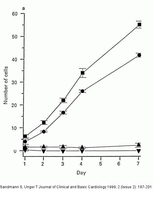
Figure/Graphic 6: Kalziumkanal - Mibefradil Inhibition of proliferation of CPAE cells by mibefradil. Cells were seeded in the absence (sqaure, control) and the presence of (circle) 1 microM, (triangle, peak up) 10 micorM and (triangle, peak down) 100 microM mibefradil. Inhibition of proliferation was significant at all concentrations. At 100 microM no living cells were present, at 10 microM adhesive cells could still be counted indicating true antiproliferative effects. Plotted are mean +/- SEM (vertical lines) at each day after seeding. |

Figure/Graphic 6: Kalziumkanal - Mibefradil
Inhibition of proliferation of CPAE cells by mibefradil. Cells were seeded in the absence (sqaure, control) and the presence of (circle) 1 microM, (triangle, peak up) 10 micorM and (triangle, peak down) 100 microM mibefradil. Inhibition of proliferation was significant at all concentrations. At 100 microM no living cells were present, at 10 microM adhesive cells could still be counted indicating true antiproliferative effects. Plotted are mean +/- SEM (vertical lines) at each day after seeding. |


