Uckermann O, Galli R, Mackenroth L, Geiger K, Steiner G, Koch E, Schackert G, Kirsch M Optical Biochemical Imaging: Potential New Applications in Neuro-Oncology European Association of NeuroOncology Magazine 2014; 4 (1): 20-26 PDF Summary Overview
| ||||||||||
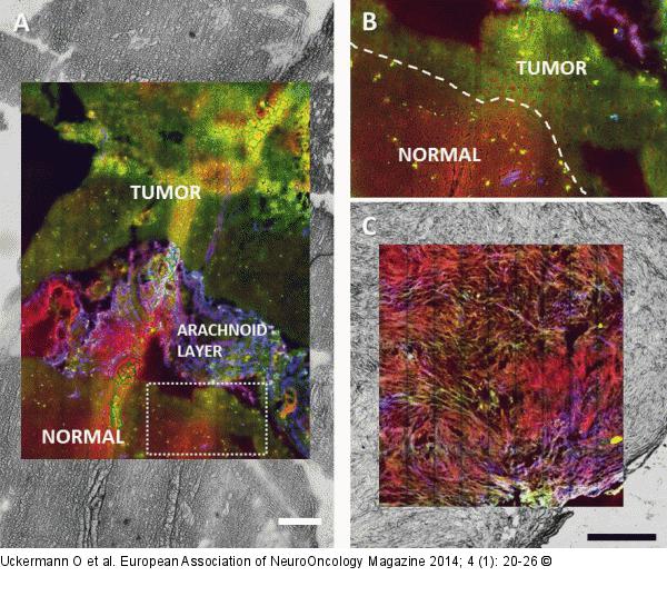
Figure/Graphic 5a-c: NLO imaging Multimodal NLO imaging of human tumours (red: CARS, green: TPEF, blue: SHG), overlaid with the bright field images of the unstained sample. The technique allows to retrieve detailed morphochemical information about tissue structure and properties on unstained samples. (a) Cryosection of human glioblastoma (scale bar: 200 μm). (b) High magnification of the border between tumour and normal tissue as indicated in (a). (c) Cryosection of human neuroma (scale bar: 0.5 mm). |

Figure/Graphic 5a-c: NLO imaging
Multimodal NLO imaging of human tumours (red: CARS, green: TPEF, blue: SHG), overlaid with the bright field images of the unstained sample. The technique allows to retrieve detailed morphochemical information about tissue structure and properties on unstained samples. (a) Cryosection of human glioblastoma (scale bar: 200 μm). (b) High magnification of the border between tumour and normal tissue as indicated in (a). (c) Cryosection of human neuroma (scale bar: 0.5 mm). |


