Wicher C, Biewald G-A Left-ventricular dysfunction, heart vagus influences and angiotensin II effects after doxorubicin perfusion in isolated rat hearts Journal of Clinical and Basic Cardiology 1999; 2 (2): 259-266 PDF Summary Overview
| ||||||||||||||||
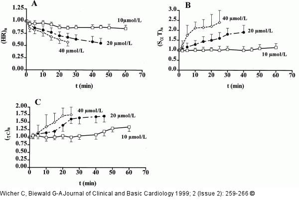
Figure/Graphic 3A-C: Doxorubicin - Angiotensin-II-Modulation Normalized changes in heart rate (HR, A), SαT (B) time of the ECG and time constant of left-ventricular contraction τC (C) during perfusion of isolated heart with doxorubicin (DXR, 10-40 micromol/L)-containing Tyrode (parameters before DXR addition were set to 1). 10 micromol/L DXR: solid line and open squares, 20 micromol/L DXR: dash-dotted line and filled circles, 40 micromol/L DXR: dashed line and open rhombs. Means and SD of 5 experiments. Note that DXR perfusion caused reductions in heart rate by up to 50 %, and increases in SαT by up to 150 %, and increase in τC by up to 75 % (after 25 minutes perfusion with 40 micromol/L DXR-containing Tyrode). |

Figure/Graphic 3A-C: Doxorubicin - Angiotensin-II-Modulation
Normalized changes in heart rate (HR, A), SαT (B) time of the ECG and time constant of left-ventricular contraction τC (C) during perfusion of isolated heart with doxorubicin (DXR, 10-40 micromol/L)-containing Tyrode (parameters before DXR addition were set to 1). 10 micromol/L DXR: solid line and open squares, 20 micromol/L DXR: dash-dotted line and filled circles, 40 micromol/L DXR: dashed line and open rhombs. Means and SD of 5 experiments. Note that DXR perfusion caused reductions in heart rate by up to 50 %, and increases in SαT by up to 150 %, and increase in τC by up to 75 % (after 25 minutes perfusion with 40 micromol/L DXR-containing Tyrode). |


