Content-type: text/html Suchergebnis - Abbildungen und Graphiken
Krause und Pachernegg
Verlag für Medizin und Wirtschaft
Mobile Version
A-  |   A  |   A+
Werbung
 
Suchergebnis - Abbildungen und Graphiken
 
Alle Journale wurden nach Smits M durchsucht Treffer 1 - 4 von 4

Medizinische Publikationen (2)  ::   Abbildungen und Graphiken  ::   Volltext (0)

Neue Suche:         Erweiterte Suche    Hilfe zur Suche

in allen Journalen nach  Bildtitel    Bildkeywords   Bildsummary   Autoren      Mindestens ein Wort  Alle Wörter   
1. Smits M
Functional Magnetic Resonance Imaging (fMRI) in Brain Tumour Patients
Brain tumour
European Association of NeuroOncology Magazine 2012; 2 (3): 123-128
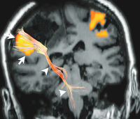
Combined fMRI and DTI tractography in a coronal view of a patient with a tumour near the primary motor cortex. Displacement of both the primary motor cortex (arrows) and the corticospinal tract (arrowheads) is seen.

Keywords: brainfMRITumor
2. Smits M
Functional Magnetic Resonance Imaging (fMRI) in Brain Tumour Patients
Brain tumour
European Association of NeuroOncology Magazine 2012; 2 (3): 123-128
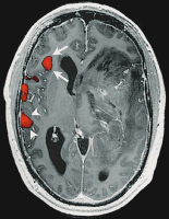
Large high-grade tumour in the left temporal lobe of a right-handed patient. fMRI activation of a verb generation task is present in the expressive-language area in the inferior frontal gyrus (arrows), as well as in the receptive-language areas in the posterior temporo-parietal cortex (arrowheads) in the right hemisphere. No activation is seen in the left hemisphere. Upon surgery, a left-lateralised hemispheric language representation was found with ECM, as would be expected in a right-handed patient. The atypical lateralisation towards the right hemisphere found with fMRI is most likely due to a tumour (mass) effect decreasing the BOLD signal in the affected left hemisphere.

Keywords: brainfMRITumor
3. Smits M
Functional Magnetic Resonance Imaging (fMRI) in Brain Tumour Patients
Brain tumour
European Association of NeuroOncology Magazine 2012; 2 (3): 123-128
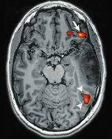
Brain tumour localised in the left temporal lobe of a left-handed patient. fMRI activation of a verb-to-noun generation is present in the expressive language area in the inferior frontal gyrus (arrow), as well as in the receptive language areas in the posterior temporo-parietal cortex (arrowheads). Activation is more pronounced in the left hemisphere, indicating a left-lateralised hemispheric language representation.

Keywords: brainfMRITumor
4. Smits M
Functional Magnetic Resonance Imaging (fMRI) in Brain Tumour Patients
Brain tumour
European Association of NeuroOncology Magazine 2012; 2 (3): 123-128
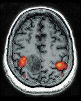
Brain tumour localised in the primary hand motor cortex as evidenced by fMRI activation of a bilateral finger tapping task adjacent to the tumour in the precentral and post-central cortex bilaterally.

Keywords: brainfMRITumor
 

 Seite 1 von 1 
 
copyright © 2000–2026 Krause & Pachernegg GmbH | Sitemap | Datenschutz | Impressum
 
Werbung