|
|
|---|
1. Wolf H;
Neuhauser C;
Franta C;
Hauer D;
Ungersböck K;
Oberndorfer S
Komplikationen bei intrathekaler Baclofentherapie
Spinaler Katheter
Journal für Neurologie, Neurochirurgie und Psychiatrie 2016; 17 (1): 13-15
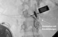
Durchleuchtung mit Darstellung des spinalen Katheters nach Kontrastmittelapplikation. Keywords: Durchleuchtung,
intrathekale Baclofentherapie,
Röntgen,
spinaler Katheter
|
|
|
|
|
|
|
|
|
4. Oberndorfer S;
Ungersböck K;
Moser W;
Riedlberger U;
Marhold F;
Heinz G;
Sedivy R;
Brustbauer R;
Kopetzky G;
Vyhnalik M;
Balcke P
Interdisziplinäre neuroonkologische Betreuung eines Patienten mit sekundärem Glioblastom
Glioblastom
Tumorboard 2014; 3 (2): 13-16
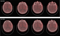
Follow-up-Imaging vom Juni 2014: im FET-PET trotz rezidivierender fokal komplexer Anfälle kein vermehrter Tracer-Uptake im Vergleich zu den Vorbildern vom Mai 2014 Keywords: FET-PET,
Glioblastom,
Onkologie
|
|
|
5. Oberndorfer S;
Ungersböck K;
Moser W;
Riedlberger U;
Marhold F;
Heinz G;
Sedivy R;
Brustbauer R;
Kopetzky G;
Vyhnalik M;
Balcke P
Interdisziplinäre neuroonkologische Betreuung eines Patienten mit sekundärem Glioblastom
Glioblastom
Tumorboard 2014; 3 (2): 13-16
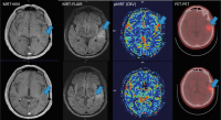
Follow-up-Imaging vom Mai 2014: MRT + KM, MRT-FLAIR und pMRT, sowie FET-PET zeigen nach wie vor keine eindeutigen Hinweise auf Tumoraktivität. Vergleiche Abb. 2 und 3 (Tumorbereich mit Pfeil markiert) Keywords: FET-PET,
FLAIR,
Glioblastom,
MRT,
Onkologie
|
|
|
6. Oberndorfer S;
Ungersböck K;
Moser W;
Riedlberger U;
Marhold F;
Heinz G;
Sedivy R;
Brustbauer R;
Kopetzky G;
Vyhnalik M;
Balcke P
Interdisziplinäre neuroonkologische Betreuung eines Patienten mit sekundärem Glioblastom
Glioblastom
Tumorboard 2014; 3 (2): 13-16
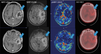
Follow-up-Imaging vom Februar 2014: MRT + KM, MRT FLAIR und pMRT, sowie FET-PET zeigen deutliche Hinweise für „Pseudoprogression“. Deutlich ausgeprägte KM-Aufnahme (MRT + KM), verminderte Perfusion in der pMRT und herabgesetzte Traceraufnahme im FET-PET im Vergleich zum Vorbefund (Tumorbereich mit Pfeil markiert) Keywords: FET-PET,
FLAIR,
Glioblastom,
MRT,
Onkologie
|
|
|
7. Oberndorfer S;
Ungersböck K;
Moser W;
Riedlberger U;
Marhold F;
Heinz G;
Sedivy R;
Brustbauer R;
Kopetzky G;
Vyhnalik M;
Balcke P
Interdisziplinäre neuroonkologische Betreuung eines Patienten mit sekundärem Glioblastom
Glioblastom
Tumorboard 2014; 3 (2): 13-16
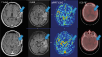
MRT (KM/FLAIR/pMRT) und FET-PET vom September 2013. Leichte KM-Aufnahme, zunehmende T2-hyperintense Läsion, sowie beginnende Hyperperfusion in der pMRT (CBV), als indirekte Zeichen einer Malignisierung. Ebenso in der FET-PET-Untersuchung vermehrter Tracer-Uptake links-temporal (Tumorbereich mit Pfeil markiert) Keywords: FET-PET,
FLAIR,
Glioblastom,
MRT,
Onkologie
|
|
|
8. Oberndorfer S;
Ungersböck K;
Moser W;
Riedlberger U;
Marhold F;
Heinz G;
Sedivy R;
Brustbauer R;
Kopetzky G;
Vyhnalik M;
Balcke P
Interdisziplinäre neuroonkologische Betreuung eines Patienten mit sekundärem Glioblastom
Glioblastom
Tumorboard 2014; 3 (2): 13-16
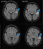
MRT/FLAIR vom Juni 2011 (radiologische Erstdiagnose) und präbioptisch im Mai 2012: zunehmende hyperintense Signalalteration links temporal (blauer Pfeil) Keywords: FLAIR,
Glioblastom,
hyperintense Signalalteration,
MRT,
Onkologie
|
|
|
9. Moser W;
Marhold F;
Ungersböck K;
Oberndorfer S
Subacute Spinal Cord Cempression Due to an Intramedullar Spinal Teratoma in a 53-Year-Old Female Patient: A Case Report
Spinal Teratoma
European Association of NeuroOncology Magazine 2014; 4 (1): 34-36
MRI of the thoracic and lumbar spine after partial resection. Keywords: MRI,
spinal teratoma
|
|
|
10. Moser W;
Marhold F;
Ungersböck K;
Oberndorfer S
Subacute Spinal Cord Cempression Due to an Intramedullar Spinal Teratoma in a 53-Year-Old Female Patient: A Case Report
Spinal Teratoma
European Association of NeuroOncology Magazine 2014; 4 (1): 34-36
Intraoperative pictures: (a) after opening of the dura mater; (b) pus-like liquid appearing during
resection of the tumour; (c) collapse of the myelon
after resection. Keywords: intraoperative picture,
photograph,
spinal teratoma
|
|
|
11. Moser W;
Marhold F;
Ungersböck K;
Oberndorfer S
Subacute Spinal Cord Cempression Due to an Intramedullar Spinal Teratoma in a 53-Year-Old Female Patient: A Case Report
Spinal Teratoma
European Association of NeuroOncology Magazine 2014; 4 (1): 34-36
MRI of the thoracic and lumbar spine at admission, showing the teratoma at levels Th11/Th12. Keywords: MRI,
spinal teratoma
|
|
|
12. Oberndorfer S
Tumorboards stellen sich vor: Neuroonkologisches Tumorboard
Neuroonkologische Tumorboard
Tumorboard 2014; 3 (1): 21-23
Zusammensetzung eines neuroonkologischen Tumorboards. Keywords: neuroonkologisches Tumorboard,
Onkologie,
Schema,
Tumorboard
|
|
|
13. Wohlgenannt V;
Oberndorfer S;
Grisold W
Intractable Headache in a Glioblastoma Patient
Glioblastoma
European Association of NeuroOncology Magazine 2012; 2 (3): 134-135
T1-weighted MRI (axial) with contrast media, showing enhancement of
the meninges (arrows), and only little enhancement at the primary tumour location
at the left temporobasal area (asterisk). Keywords: glioblastoma,
MRI
|
|
|
14. Wohlgenannt V;
Oberndorfer S;
Grisold W
Intractable Headache in a Glioblastoma Patient
Glioblastoma
European Association of NeuroOncology Magazine 2012; 2 (3): 134-135
T1-weighted MRI (sagittal) with contrast media, showing enhancement
of the meninges (arrows). Keywords: glioblastoma,
MRI
|
|
|
15. Grisold W;
Oberndorfer S;
Windebank AJ
Chemotherapy and Polyneuropathies
Nerve Roots
European Association of NeuroOncology Magazine 2012; 2 (1): 25-36
Autopsy of the nerve
roots, cauda equina and posterior
columns of a 74-year-old woman with
breast cancer and suspected meningeal
carcinomatosis. She received
several cycles of intrathecal cytosine
arabinoside (Depo-Cyt®). The
myelin appears blue with a Klüver
staining. (a) Cauda equina. The
nerve roots show loss of myelin with
a patchy damage to nerve fascicles.
(b) Spinal thoracic nerve. Diffuse
demyelination. (c) Spinal cord and
posterior column: Diffuse loss of
myelinated fibres, ballooned degeneration
of myelinated fibres. Keywords: cauda equina,
nerve roots,
posterior column,
spinac thoracic nerve,
spinal cord
|
|
|
16. Grisold W;
Oberndorfer S;
Windebank AJ
Chemotherapy and Polyneuropathies
Cisplatinum neuropathy
European Association of NeuroOncology Magazine 2012; 2 (1): 25-36
Cisplatinum neuropathy. Clinical and electrophysiological features of a
40-year-old male with an ataxic cisplatinum neuropathy. (a) Despite numbness and
sensory ataxia no motor involvement with normal small-hand muscles. Nerve conduction
velocity (NCV) measurement shows normal motor NCV, (b) the sensory NCV is
reduced to 1 uV (pathologic). (c) The feet appear normal, neither atrophy nor trophic
changes can be seen. (d) Knee-to-shin is pathologic, vibration perception is absent.
(e) Motor NCVs of peroneal nerves are normal. The sural nerve sensory potential is
absent; (f) the F wave is normal. Keywords: ataxic cisplatinum neuropathy,
clinical features
|
|
|
17. Grisold W;
Oberndorfer S;
Windebank AJ
Chemotherapy and Polyneuropathies
CIPN
European Association of NeuroOncology Magazine 2012; 2 (1): 25-36
Morphologic correlates of CIPN. The DRGs can be the target of cumulative
neurotoxicity as in platinum compounds. Axons, rarely the myelin, can be damaged.
At the neuromuscular transmission site (NMT), the acute toxicity of oxaliplatin occurs.
Unmyelinated fibers and terminal nerve arbors are the sites of acute taxane
toxicity. An additional spinal mechanism at the alpha-2-delta-Typ1 subunit in neuropathic
pain is assumed. Keywords: CIPN,
scheme
|
|
|
18. Lindeck-Pozza E;
Oberndorfer S;
Hainfellner JA;
Grisold W
Paraneoplastische neurologische Syndrome
Paraneoplstische neurologische Syndrome
Journal für Neurologie, Neurochirurgie und Psychiatrie 2009; 10 (2): 26-31
Flussdiagramm zur Diagnostik paraneoplstischer neurologischer Syndrome. Mod. nach [1]. Keywords: Neurologie
|
|
|
19. Pakrah-Bodingbauer B;
Loyoddin M;
Oberndorfer S;
Kleinpeter G
Wertigkeit der 5-Aminolävulinsäure- (5-ALA-) gestützten Gliomchirurgie
Tumor
Journal für Neurologie, Neurochirurgie und Psychiatrie 2009; 10 (2): 22-25
Links: OP-Situs unter Weißlicht; Mitte: 5-ALA wird von malignen Gliomen in eine fluoreszierende Substanz umgewandelt; rechts: Darstellung von Resttumor unter Fluoreszenzlicht Keywords: 5-ALA,
Tumor
|
|
|
20. Pakrah-Bodingbauer B;
Loyoddin M;
Oberndorfer S;
Kleinpeter G
Wertigkeit der 5-Aminolävulinsäure- (5-ALA-) gestützten Gliomchirurgie
5-ALA
Journal für Neurologie, Neurochirurgie und Psychiatrie 2009; 10 (2): 22-25
Umstellung des Mikroskoplichts auf Blaulicht zur Anwendung von 5-ALA Keywords: 5-ALA,
Foto,
Operationssitus
|
|
|
21. Pakrah-Bodingbauer B;
Loyoddin M;
Oberndorfer S;
Kleinpeter G
Wertigkeit der 5-Aminolävulinsäure- (5-ALA-) gestützten Gliomchirurgie
Tumor
Journal für Neurologie, Neurochirurgie und Psychiatrie 2009; 10 (2): 22-25
Neuronavigierte Darstellung der Tumors, OP-Situs unter normalem Mikroskoplicht Keywords: 5-ALA,
Tumor
|
|
|
22. Oberndorfer S;
Grisold W
Lumbosakrale Plexopathien bei Tumorpatienten
Lumosakrale Plexopathie
Journal für Neurologie, Neurochirurgie und Psychiatrie 2004; 5 (1): 14-16
CT-Abdomen mit tumoröser Auftreibung des linken M. iliopsoas (weißer Pfeil). Keywords: Computertomographie,
Musculus iliopsoas,
Neurologie,
Plexopathie,
Plexus lumbosacralis,
Tumor
|
|
|
| |

 Suche Medizinische Publikationen
 Suche Abbildungen und Grafiken
 Medizinprodukte - Labortechnik
 Antidotarium (Stand 2012)
 Phytokodex (Stand 2012)
 Fachbücher
 Patientenliteratur