|
|
|---|
1. Montag M;
Toth B;
Strowitzki T
New Laboratory Techniques in Reproductive Medicine
Series of time-lapse images
Journal für Reproduktionsmedizin und Endokrinologie - Journal of Reproductive Medicine and Endocrinology 2013; 10 (Sonderheft 1): 33-37
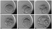
Series of time-lapse images taken from one embryo starting at the 2PN stage (upper left) up to the expanded blastocyst stage (lower right). Images were taken at 19.1, 33.1, 47.1, 70.3, 89.3 and 114.1 hours after ICSI and following transfer this embryo implanted and gave a pregnancy. Analysis of cleavage times and intervals between cleavages allows for subtle differentiation between individual embryos and helps choosing the one with the most synchronous and timed development. Keywords: 2PN stage,
blastocyst stage,
time-lapse
|
|
|
2. Montag M;
Toth B;
Strowitzki T
New Laboratory Techniques in Reproductive Medicine
Result of an array-CGH analysis
Journal für Reproduktionsmedizin und Endokrinologie - Journal of Reproductive Medicine and Endocrinology 2013; 10 (Sonderheft 1): 33-37
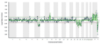
This example shows the result of an array-CGH analysis of the 1st polar body from an oocyte taken from a patient with a maternal reciprocal translocation 46,XX,t(5;8)(q22;p11.2). The possibility to detect numerical (loss of a chromatid 15 and 16) and structural chromosomal aberrations (gain of a chromatid 8p and loss of a chromatid 5q) by array-CGH is obvious. Reprinted in part with permission from [ Montag et al., J Reproduktionsmed Endokrinol 2010; 6: 498–502]. Keywords: array-CGH
|
|
|
3. Montag M;
Toth B;
Strowitzki T
Sperm Selection in ART
Sperm selection
Journal für Reproduktionsmedizin und Endokrinologie - Journal of Reproductive Medicine and Endocrinology 2012; 9 (6): 485-489
Sperm selection by hyaluronic acid binding.
The figure shows a dish where the lower right part is
coated with hyaluronic acid. Sperm with hyaluronic
acid binding sites attach to the coated part and are expected
to be mature and suitable for ICSI. Spermatozoa
at the upper left corner have not bound and are still
motile. These sperm are immature and should not be
used for ICSI. Keywords: hyaluronic acid binding,
ICSI,
sperm
|
|
|
4. Montag M;
Toth B;
Strowitzki T
Sperm Selection in ART
Sperm
Journal für Reproduktionsmedizin und Endokrinologie - Journal of Reproductive Medicine and Endocrinology 2012; 9 (6): 485-489
Comparison of digital enhanced high magnification
image versus conventional Hoffman modulation
contrast. This figure shows a picture of two sperm
taken with a standard Hofmann 40× objective (A) and a
63× objective with a high numerical aperture and using
Cytoscreen (B) a digital image enhancement software
(Octax, Bruckberg, Germany). The presence of a small
vacuole in the head of the right sperm as visualized by
digital enhanced high magnification can be seen in the
standard Hofmann contrast picture as a irregularity in
the sperm head. Reprinted from [23]. Keywords: sperm,
vacuole
|
|
|
5. Montag M;
Toth B;
Strowitzki T
Sperm Selection in ART
Sperm viability
Journal für Reproduktionsmedizin und Endokrinologie - Journal of Reproductive Medicine and Endocrinology 2012; 9 (6): 485-489
Sperm viability as proven by laser. A viable
sperm is shown after the application of a single laser
shot. The laser shot caused a curling of the end of the
sperm tail which is indicative for sperm viability. Nonviable
sperm show no reaction of the sperm tail. Despite
the curling of the sperm tail these sperm can be
used for ICSI without any further treatment as the laser
causes also a permeabilization of the sperm plasma
membrane [15]. Keywords: single laser shot,
sperm
|
|
|
|
|
|
7. Montag M;
Köster K;
van der Ven K;
Bohlen U;
Bender F;
van der Ven H
Kombinierte Translokations- und Aneuploidieuntersuchungen nach Polkörperbiopsie und array-Comparative Genomic Hybridisation
Translokationen
Journal für Reproduktionsmedizin und Endokrinologie - Journal of Reproductive Medicine and Endocrinology 2010; 7 (6): 498-502
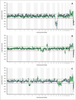
Scan-Auswertungsbeispiele für die 3 vorgestellten Translokationen. (A): Verlust zweier Chromatiden 14 im 2. Polkörper einer Eizelle bei Vorliegen einer balancierten
mütterlichen Robertson’schen Translokation 45,XX,rob(13;14)(q10;q10). (B): Zugewinn einer Chromatide 11p und Verlust einer Chromosoms 19p sowie einer Chromatide für
Chromosom 9 im 1. Polkörper bei Vorliegen einer balancierten mütterlichen reziproken Translokation 46,XX,t(11;19)(p10;p10). (C): Zugewinn einer Chromatide 8p und Verlust einer
Chromatide 5q sowie je einer Chromatide für die Chromosom 15 und 16 im 1. Polkörper bei Vorliegen einer balancierten mütterlichen reziproken Translokation 46,XX,t(5;8)(q22;p11.2).
A: Robertson’sche Translokation 45,XX,rob(13;14)(q10;q10) B: Reziproke Translokation 46,XX,t(11;19)(p10;p10) C: Reziproke Translokation 46,XX,t(5;8)(q22;p11.2) Keywords: Biologie,
Embryologie,
Genetik,
Scan,
Translokation
|
|
|
8. Montag M;
Schwarz C;
Köster M;
van der Ven K;
van der Ven H
IMSI (intracytoplasmic morphologically selected sperm injection): Therapieoptimierung bei ausgeprägter Störung der Spermatogene?
Vakuolen
Journal für Reproduktionsmedizin und Endokrinologie - Journal of Reproductive Medicine and Endocrinology 2009; 6 (4): 153-155
Darstellung von Vakuolen im Spermienkopf
an einem gut justierten Standard-ICSI-Mikroskop
(Leica DMIRB), ausgestattet mit einem 40× Hofmann-Kontrast-Objektiv (A) und an einem ICSI-Mikroskop (Nikon TE2000), ausgestattet mit einem 60× Differenziellen
Interferenz-Kontrast-Objektiv (B) mit hoher numerischer Aperatur und Bildbearbeitung (CytoScreen, Octax). Die Pfeile weisen auf Vakuolen im Spermienkopf hin, deren Präsenz auch an dem Standard-ICSI-Mikroskop
bereits gut erkannt werden kann. Zur Vergleichbarkeit wurden beide Abbildungen auf die gleiche Endvergrößerung
eingestellt. Keywords: Endokrinologie,
Gynäkologie,
Mikroskopische Aufnahme,
Spermienkopf,
Vakuole
|
|
|
9. Montag M;
Köster K;
van der Ven K;
van der Ven H
Qualität der Eizelle
Eizelle
Journal für Reproduktionsmedizin und Endokrinologie - Journal of Reproductive Medicine and Endocrinology 2009; 6 (Sonderheft 1): 26-28
Qualitätsbeurteilung
einer menschlichen Eizelle.
A: Bei der dargestellten Eizelle können im normalen mikroskopischen Bild 2 Polkörper (Pfeile) ausgemacht werden. B: Die polarisationsmikroskopische Beurteilung
zeigt, dass in unmittelbarer Nähe zu jedem Polkörper eine Metaphase-II-Spindel vorliegt
und es sich folglich um eine diploide
Eizelle handelt. Keywords: Eizelle,
Polarisationsmikroskop,
Reproduktionsmedizin
|
|
|
|
|
|
|
|
|
12. Montag M;
van der Ven H;
van der Ven K
Erste klinische Erfahrungen mit der Polkörperdiagnostik in Deutschland
Chromosomen-Fehlverteilungen
Journal für Fertilität und Reproduktion 2002; 12 (4) (Ausgabe für Schweiz): 4-8
Journal für Fertilität und Reproduktion 2002; 12 (4) (Ausgabe für Österreich): 7-12
Chromosomen-Fehlverteilungen in Eizellen älterer Patientinnen nach Untersuchung der Chromosomen 13, 16, 18, 21 und 22 Keywords: Chromosomenaberration,
Diagramm,
Fertilität,
Monosomie,
Trisomie,
ältere Patientin
|
|
|
|
|
|
14. Montag M;
van der Ven H;
van der Ven K
Erste klinische Erfahrungen mit der Polkörperdiagnostik in Deutschland
Polkörperentnahme
Journal für Fertilität und Reproduktion 2002; 12 (4) (Ausgabe für Schweiz): 4-8
Journal für Fertilität und Reproduktion 2002; 12 (4) (Ausgabe für Österreich): 7-12
Zur Entnahme des 1. Polkörpers wird die Zona pellucida einer menschlichen Eizelle mit einem Diodenlaser direkt über dem 1. Polkörper eröffnet (Abb. 1A). Der Polkörper wird in eine stumpfe, feuerpolierte Biopsienadel aspiriert und steht für weitere Untersuchungen zur Verfügung (Abb. 1B) Keywords: Eizelle,
Fertilität,
Foto,
Polkörperentnahme
|
|
|
| |

 Suche Medizinische Publikationen
 Suche Abbildungen und Grafiken
 Medizinprodukte - Labortechnik
 Antidotarium (Stand 2012)
 Phytokodex (Stand 2012)
 Fachbücher
 Patientenliteratur