| |
| Alle Journale wurden nach durchsucht |
Treffer 11441 - 11480 von 11970 |
|
|
|---|
11441. Jarius Ch;
Budka H
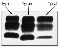
Übertragbare spongiforme Enzephalopathien
Spongiforme Enzephalopathie - Prionprotein-Glykoformen
Journal für Neurologie, Neurochirurgie und Psychiatrie 2002; 3 (2): 7-17
Western Blot: nach Auftrennung proteaseresistenter Fraktionen des Prionproteins können 3 Glykoformen des Prionproteins unterschieden werden (Typ 1, Typ 2A und Typ 2B) Keywords: Glykoform,
Neurologie,
Prionprotein,
Western Blot
|
|
|
11442. Jarius Ch;
Budka H
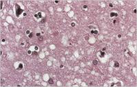
Übertragbare spongiforme Enzephalopathien
Spongiforme Enzephalopathie
Journal für Neurologie, Neurochirurgie und Psychiatrie 2002; 3 (2): 7-17
Typische spongiforme Gewebealteration in der Großhirnrinde bei CJK (Hämatoxylin-Eosin-Fbg.) Keywords: Cortex cerebri,
Großhirnrinde,
Histologisches Präparat,
Neurologie,
Spongiforme Enzephalopathie
|
|
|
11443. Bocker B;
Smolenski UC
Physikalische Therapie der Beckenbodeninsuffizienz - Methodenvergleich
Beckenbodeninsuffizienz
Journal für Urologie und Urogynäkologie 2002; 9 (2) (Ausgabe für Schweiz): 16-22
Journal für Urologie und Urogynäkologie 2002; 9 (2) (Ausgabe für Österreich): 20-27
Veränderung der Bewertungsparameter während und nach Behandlung mit dem indikationsabhängigen Therapiemittel der Wahl (n = 2 x 24, MV) Keywords: Beckenbodeninsuffizienz,
Biofeedback,
Diagramm,
Elektrostimulation,
Inkontinenzepisoden,
Urogynäkologie,
Urologie
|
|
|
11444. Bocker B;
Smolenski UC
Physikalische Therapie der Beckenbodeninsuffizienz - Methodenvergleich
Beckenbodeninsuffizienz
Journal für Urologie und Urogynäkologie 2002; 9 (2) (Ausgabe für Schweiz): 16-22
Journal für Urologie und Urogynäkologie 2002; 9 (2) (Ausgabe für Österreich): 20-27
Diagnosenabhängige Änderung des Harninkontinenzgrades während und nach EMG-getriggerter Elektrostimulation (n = 2 x 24, MV, CI) Keywords: Beckenbodeninsuffizienz,
Diagramm,
Elektrostimulation,
Harninkontinenzgrad,
Urogynäkologie,
Urologie
|
|
|
11445. Bocker B;
Smolenski UC
Physikalische Therapie der Beckenbodeninsuffizienz - Methodenvergleich
Beckenbodeninsuffizienz
Journal für Urologie und Urogynäkologie 2002; 9 (2) (Ausgabe für Schweiz): 16-22
Journal für Urologie und Urogynäkologie 2002; 9 (2) (Ausgabe für Österreich): 20-27
Diagnosenabhängige Änderung des Harninkontinenzgrades während und nach Biofeedbacktherapie (n = 2 x 24, MV, CI) Keywords: Beckenbodeninsuffizienz,
Biofeedback,
Diagramm,
Harninkontinenzgrad,
Urogynäkologie,
Urologie
|
|
|
|
|
|
|
|
|
|
|
|
11449. Steiner T;
Braunsdorf A;
Burkhardt F;
Janitzky V;
Junker K;
Schubert J
Genetische Veränderungen an primären Prostatakarzinom-Biopsien in Korrelation zum klinischen Verlauf
Vergleichende genomische Hybridisierung
Journal für Urologie und Urogynäkologie 2002; 9 (2) (Ausgabe für Schweiz): 5-9
Journal für Urologie und Urogynäkologie 2002; 9 (2) (Ausgabe für Österreich): 7-11
CGH-Profil eines Patienten, welcher im Nachbeobachtungszeitraum von 36 Monaten einen Tumorprogreß entwickelte; beachte den Zugewinn 8q Keywords: Andrologie,
CGH,
Diagramm,
Urologie,
Vergleichende genomische Hybridisierung
|
|
|
|
|
|
11451. Raufi A;
Khan IA;
Nair VM;
Rahmatullah SI;
Rodriguez C;
Sacchi TJ;
Sahni G;
Vasavada BC
Rupture of a Surgically Repaired Sinus of Valsalva Aneurysm
Sinus-Valsalvae-Aneurysma
Journal of Clinical and Basic Cardiology 2002; 5 (2): 199-200
Aortogram demonstrates a communication between aorta and right atrium (RA) Keywords: Aneurysma,
Aneurysma,
Aorta,
Aorta,
Aortogramm,
Aortogramm,
Atrium,
Atrium,
Shunt,
Shunt,
Sinus valsalvae,
Sinus valsalvae
|
|
|
11452. Raufi A;
Khan IA;
Nair VM;
Rahmatullah SI;
Rodriguez C;
Sacchi TJ;
Sahni G;
Vasavada BC
Rupture of a Surgically Repaired Sinus of Valsalva Aneurysm
Sinus-Valsalvae-Aneurysma
Journal of Clinical and Basic Cardiology 2002; 5 (2): 199-200
Transoesophageal echocardiography demonstrates dilated right atrium (RA) and right ventricle (RV) and a high velocity shunt from aorta to right atrium. LA = left atrium, LV = left ventricle Keywords: Aneurysma,
Aneurysma,
Aorta,
Aorta,
Atrium,
Atrium,
echocardiography,
Echokardiographie,
Ruptur,
rupture,
Shunt,
Shunt,
Sinus valsalvae,
Sinus valsalvae,
TEE,
TEE,
ventricle,
Ventrikel
|
|
|
11453. Krayenbuehl P-A;
Christen S
Tricyclic Antidepressant Overdose Mimicking Myocardial Infarction
EKG - Trizyklika - Myokardinfarkt
Journal of Clinical and Basic Cardiology 2002; 5 (2): 197
Serial standard electrocardiographic tracings during hospitalization Keywords: Antidepressivum,
EKG,
electrocardiography,
Elektrokardiographie,
Mimikry,
myocardial infarction,
Myokardinfarkt,
Overdose,
Tricyclic antidepressant,
Trizyklikum,
Überdosis
|
|
|
11454. Dahm JB;
Hummel A;
Kuon E;
Voelzke H;
Vogelgesang D
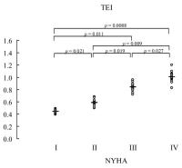
Doppler Echocardiography in Dilated Cardiomyopathy: Diastolic and Combined Systolic/Diastolic Parameters Offer More Detailed Information on Left Ventricular Global Dysfunction than Systolic Parameters
Systolisch/diastolische Performance
Journal of Clinical and Basic Cardiology 2002; 5 (2): 189-192
The systolic/diastolic performance (Tei-index) of the 68 patients in the different NYHA-groups Keywords: Diagramm,
diastole,
diastole,
NYHA,
NYHA,
Performance,
Systole,
Systole,
TEI-Index,
TEI-Index
|
|
|
11455. Dahm JB;
Hummel A;
Kuon E;
Voelzke H;
Vogelgesang D
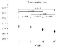
Doppler Echocardiography in Dilated Cardiomyopathy: Diastolic and Combined Systolic/Diastolic Parameters Offer More Detailed Information on Left Ventricular Global Dysfunction than Systolic Parameters
A-Dezelerationszeit
Journal of Clinical and Basic Cardiology 2002; 5 (2): 189-192
A-deceleration time of the 68 patients in the different NYHA-groups. Keywords: A-deceleration time,
A-Dezeleration,
Diagramm,
NYHA,
NYHA
|
|
|
11456. Dahm JB;
Hummel A;
Kuon E;
Voelzke H;
Vogelgesang D
Doppler Echocardiography in Dilated Cardiomyopathy: Diastolic and Combined Systolic/Diastolic Parameters Offer More Detailed Information on Left Ventricular Global Dysfunction than Systolic Parameters
E-Dezelerationszeit
Journal of Clinical and Basic Cardiology 2002; 5 (2): 189-192
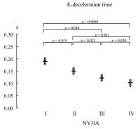
E-deceleration time of the 68 patients in the different NYHA-groups. Keywords: Diagramm,
E-deceleration time,
E-Dezeleration,
NYHA,
NYHA
|
|
|
11457. Dahm JB;
Hummel A;
Kuon E;
Voelzke H;
Vogelgesang D
Doppler Echocardiography in Dilated Cardiomyopathy: Diastolic and Combined Systolic/Diastolic Parameters Offer More Detailed Information on Left Ventricular Global Dysfunction than Systolic Parameters
LVEF
Journal of Clinical and Basic Cardiology 2002; 5 (2): 189-192
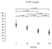
Left ventricular ejection fraction (LV-EF) obtained by angiography of the 68 patients in the different NYHA-groups. Keywords: Angiographie,
angiography,
Auswurffraktion,
Ejection fraction,
Ejektion,
NYHA,
NYHA,
ventricle,
Ventrikel
|
|
|
11458. Dahm JB;
Hummel A;
Kuon E;
Voelzke H;
Vogelgesang D
Doppler Echocardiography in Dilated Cardiomyopathy: Diastolic and Combined Systolic/Diastolic Parameters Offer More Detailed Information on Left Ventricular Global Dysfunction than Systolic Parameters
LVEF
Journal of Clinical and Basic Cardiology 2002; 5 (2): 189-192
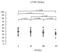
Left ventricular ejection fraction (LV-EF) obtained by
echocardiography (Simpson’s method) of the 68 patients in the different NYHA-groups. Keywords: Auswurffraktion,
Diagramm,
echocardiography,
Echokardiographie,
Ejection fraction,
Ejektion,
LVEF,
NYHA,
NYHA,
Simpson,
Simpson,
ventricle,
Ventrikel
|
|
|
11459. Dahm JB;
Hummel A;
Kuon E;
Voelzke H;
Vogelgesang D
Doppler Echocardiography in Dilated Cardiomyopathy: Diastolic and Combined Systolic/Diastolic Parameters Offer More Detailed Information on Left Ventricular Global Dysfunction than Systolic Parameters
LVEDD
Journal of Clinical and Basic Cardiology 2002; 5 (2): 189-192
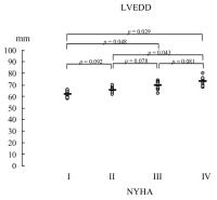
Left ventricular enddiastolic diameter (LVEDD) measurements obtained by echocardiography of the 68 patients in the different NYHA-groups. Keywords: Diagramm,
Diameter,
Durchmesser,
echocardiography,
Echokardiographie,
LVEDD,
LVEDD,
NYHA,
NYHA
|
|
|
11460. Dahm JB;
Hummel A;
Kuon E;
Voelzke H;
Vogelgesang D
Doppler Echocardiography in Dilated Cardiomyopathy: Diastolic and Combined Systolic/Diastolic Parameters Offer More Detailed Information on Left Ventricular Global Dysfunction than Systolic Parameters
Systolisch/diastolische Performance
Journal of Clinical and Basic Cardiology 2002; 5 (2): 189-192
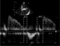
Calculation of the systolic/diastolic performance
[(ICT+IRT)/ET]: Mitral closure-to-opening interval (a) is characterized as the time from cessation to the beginning of mitral inflow. Ejection time (ET) is measured as the duration of left ventricular outflow (b). Isovolumetric contraction time (ICT) + isovolumetric relaxation time (IRT) is calculated by subtracting "b" from "a" divided by "b" [(ICT+IRT)/ET = (a–b)/b]. Keywords: diastole,
diastole,
Echocardiographa,
Echokardiographie,
Systole,
Systole
|
|
|
11461. Sperker W;
Glogar HD;
Gyöngyösi M
Percutaneous Treatment of Left Main Coronary Artery Stenoses
Koronararterienintervention
Journal of Clinical and Basic Cardiology 2002; 5 (2): 163-169
Angiograms of an unprotected left main coronary artery intervention and of the 6-month follow-up (6-mo FUP) result Keywords: Angiographie,
angiography,
Arteria coronaria sinistra,
Arteria coronaria sinistra,
Intervention,
Intervention,
Stent,
Stent
|
|
|
11462. Brainin M
Statins and Stroke: A Promising Approach Towards Stroke Prevention
Zerebrale Blutung
Journal of Clinical and Basic Cardiology 2002; 5 (2): 159-162
MR scans of a 74-year old patient with amyloidangiopathyinduced cerebral bleeding in the right parietal subcortex. In the haemosensitive scans (gradient-echo T2* sequences) numerous
additional hypointense lesions are visible, indicating multiple silent cerebral "microbleeds". Such patients might bear an additional risk for a recurrent haemorrhage if cholesterol or LDL is markedly reduced below normal. Keywords: Amyloidangiopathie,
amyloidangiopathy,
Bleeding,
Blutung,
Lesion,
Läsion,
Magnetic Resonance Tomography,
Magnetresonanztomographie,
Microbleeds,
Mikroblutung,
Subkortex,
Subkortex
|
|
|
11463. Scholten Ch;
Baumgartner H
Hand Carried Echocardiography Systems - A Beautiful Toy or a Useful Device With Major Impact on Clinical Practice?
Tragbares Ultraschallgerät
Journal of Clinical and Basic Cardiology 2002; 5 (2): 153-154
SonoHeart (TM) System, SonoSite, Inc., Bothwell, WA Keywords: Echokardiographie,
Gerät,
SonoHeart,
SonoSite
|
|
|
11464. Binder T
Three-Dimensional Echocardiography - Principles and Promises
3D-Echokardiographie
Journal of Clinical and Basic Cardiology 2002; 5 (2): 149-152
Model of the mitral valve (in a patient with mitral stenosis) created from a 3-dimensional dataset using stereolithography. Keywords: 3D,
3D,
Echokardiographie,
Echokardiographie,
Mitral valve,
Mitralklappe,
Stenose,
Stenosis,
Stereolithographie,
Stereolithography
|
|
|
11465. Binder T
Three-Dimensional Echocardiography - Principles and Promises
3D-Echokardiographie
Journal of Clinical and Basic Cardiology 2002; 5 (2): 149-152
Three-dimensional analysis of left ventricular function allows computation of global and regional ejection fraction (bottom) and the three-dimensional display of the ventricle (top panel). Keywords: 3D,
3D,
Auswurffraktion,
echocardiography,
Echokardiographie,
Ejection fraction,
Ejektion,
Function,
Funktion,
ventricle,
Ventrikel
|
|
|
11466. Binder T
Three-Dimensional Echocardiography - Principles and Promises
3D-Echokardiographie
Journal of Clinical and Basic Cardiology 2002; 5 (2): 149-152
Dissection of the descending aorta. The true lumen is separated from the false lumen by an intima flap. Keywords: 3D,
3D,
Aorta descendens,
Aorta descendens,
dissection,
Dissektion,
echocardiography,
Echokardiographie,
Intima,
Intima
|
|
|
11467. Binder T
Three-Dimensional Echocardiography - Principles and Promises
3D-Echokardiographie
Journal of Clinical and Basic Cardiology 2002; 5 (2): 149-152
Three-dimensional reconstruction of a patient with mitral stenosis, morphologic features such as doming of the anterior leaflet and commissural fusion can be noticed. In addition, quantification of mitral valve area can be performed. Keywords: 3D,
3D,
echocardiography,
Echokardiographie,
Mitral valve,
Mitralklappe,
Stenose,
Stenosis
|
|
|
11468. Binder T
Three-Dimensional Echocardiography - Principles and Promises
3D-Echokardiographie
Journal of Clinical and Basic Cardiology 2002; 5 (2): 149-152
Patient with a partial flail leaflet as seen during mitral valve repair. 3D-Echo is able to demonstrate the location and morphology on the defect (arrow). Keywords: 3D,
3D,
echocardiography,
Echokardiographie,
Mitral valve,
Mitralklappe,
Reconstruction,
Rekonstruktion
|
|
|
11469. Binder T
Three-Dimensional Echocardiography - Principles and Promises
3D-Echokardiographie
Journal of Clinical and Basic Cardiology 2002; 5 (2): 149-152
Three-dimensional reconstruction of the mitral valve (left) as seen from the left atrium. An anatomical specimen for comparison is shown on the right. Keywords: 3D,
3D,
echocardiography,
Echokardiographie,
Mitral valve,
Mitralklappe,
Reconstruction,
Rekonstruktion
|
|
|
11470. Kuntz-Hehner St;
Becher H;
Luederitz B;
Omran H;
Schlosser Th;
Tiemann K
Assessment of Myocardial Perfusion by Contrast Echocardiography - Ready for Clinical Practice?
Kontrastechokardiographie
Journal of Clinical and Basic Cardiology 2002; 5 (2): 145-148
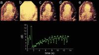
Negative bolus destruction reperfusion study using power pulse inversion imaging. First, continuous infusion of a contrast agent is adjusted to achieve full myocardial opacification using real-time imaging at very low emission power (a) (MI = 0.09). After destruction of the microbubbles by a short period of high power imaging (b) (MI = 1.3) myocardial contrast replenishment is recorded at low emission power (c–e) (MI = 0.09). A region of interest (ROI) is placed in the septum and repeated
measurements are made at the endsystolic frames. The graph shows the reperfusion curve for this ROI, fitting well to the monoexponential function. Keywords: Contrast agent,
echocardiography,
Echokardiographie,
Kontrastmittel,
reperfusion,
reperfusion
|
|
|
11471. Kuntz-Hehner St;
Becher H;
Luederitz B;
Omran H;
Schlosser Th;
Tiemann K
Assessment of Myocardial Perfusion by Contrast Echocardiography - Ready for Clinical Practice?
Kontrastechokardiographie
Journal of Clinical and Basic Cardiology 2002; 5 (2): 145-148
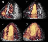
Different degrees of myocardial contrast using harmonic power Doppler imaging (four chamber view) during continuous infusion of Levovist (R): Arrival of the contrast agent in the right ventricle (a) and complete left ventricular opacification shortly after (b) using triggered imaging every heart cycle (1:1). Complete left ventricular myocardial opacification using higher trigger intervals every 3rd (c) and every 5th (d) cardiac cycle. Keywords: Contrast agent,
echocardiography,
Echokardiographie,
Four chamber view,
Kontrastmittel,
Levovist,
Levovist,
Schering GesmbH,
Schering GesmbH,
Vierkammerblick
|
|
|
11472. Rosenhek R;
Binder T
Monitoring of Invasive Procedures - The Role of Echocardiography in Cathlab and Operating Room
Echokardiographie - Kardiomyopathie
Journal of Clinical and Basic Cardiology 2002; 5 (2): 139-143
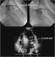
The septal branch of the left anterior descending coronary artery in a patient with hypertrophic obstructive cardiomyopathy (top panels). Estimation of the size of the septal vascular territory with myocardial contrast echocardiography (lower panel). LA = left atrium, RA = right atrium, LV = left ventricle, RV = right ventricle Keywords: Angiographie,
Arteria coronaria sinistra,
Arteria coronaria sinistra,
cardiomyopathy,
echocardiography,
Echokardiographie,
Kardiomyopathie
|
|
|
11473. Rosenhek R;
Binder T
Monitoring of Invasive Procedures - The Role of Echocardiography in Cathlab and Operating Room
Amplatzer-Occluder - Positionierung
Journal of Clinical and Basic Cardiology 2002; 5 (2): 139-143
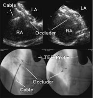
Positioning of an Amplatzer atrial septal defect occluder under TEE guidance (top left panel) and assessment of the result after release of the occluder (top right panel). The lower panels show the corresponding images obtained by fluoroscopy. LA = left atrium, RA = right atrium Keywords: Amplatzer-Occluder,
Amplatzer-Occluder,
echocardiography,
Echokardiographie,
Fluoroscopy,
Fluoroskopie,
TEE,
TEE
|
|
|
11474. Rosenhek R;
Binder T
Monitoring of Invasive Procedures - The Role of Echocardiography in Cathlab and Operating Room
Echokardiographie - Mitralklappe
Journal of Clinical and Basic Cardiology 2002; 5 (2): 139-143
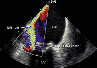
Large paravalvular leak detected by color Doppler echocardiography in a bioprosthetic mitral valve (MV-Bio Prosth). LA = left atrium, LV = left ventricle Keywords: Colordoppler,
echocardiography,
Echokardiographie,
Farbdoppler,
Mitral valve,
Mitralklappe,
Prosthesis,
Prothese
|
|
|
11475. Rosenhek R;
Binder T
Monitoring of Invasive Procedures - The Role of Echocardiography in Cathlab and Operating Room
Echokardiographie - Mitralklappen-OP
Journal of Clinical and Basic Cardiology 2002; 5 (2): 139-143
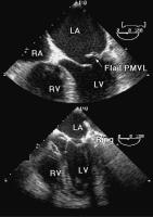
Flail posterior mitral valve leaflet (PMVL) as mechanism of mitral regurgitation (top panel). Immediate postoperative result after mitral valve reconstruction with annuloplasty ring implantation (lower panel). LA = left atrium, LV = left ventricle, RV = right ventricle Keywords: Annuloplastie,
annuloplasty,
echocardiography,
Echokardiographie,
Mitral valve,
Mitralklappe,
Reconstruction,
Rekonstruktion
|
|
|
11476. Bartel T;
Caspari G;
Erbel R;
Mueller S
Intracardiac Echocardiography - Technology and Clinical Role
Intrakardiale Echokardiographie
Journal of Clinical and Basic Cardiology 2002; 5 (2): 133-137
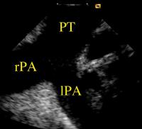
Left and right pulmonary arteries viewed with the
AcuNav (TM)-catheter in the pulmonary trunk. lPA = left pulmonary artery; rPA = right pulmonary artery; PT = pulmonary trunk Keywords: Arteria pulmonalis,
Arteria pulmonalis,
Catheter,
echocardiography,
Echokardiographie,
Katheter,
Truncus pulmonalis,
Truncus pulmonalis
|
|
|
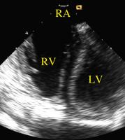
11477. Bartel T;
Caspari G;
Erbel R;
Mueller S
Intracardiac Echocardiography - Technology and Clinical Role
Intrakardiale Echokardiographie
Journal of Clinical and Basic Cardiology 2002; 5 (2): 133-137
The right ventricle viewed with the AcuNav (TM)-catheter pressed against the roof of the right atrium. LV = left ventricle; RA = right atrium; RV = right ventricle Keywords: Atrium,
Atrium,
Catheter,
echocardiography,
Echokardiographie,
Katheter,
ventricle,
Ventrikel
|
|
|
11478. Bartel T;
Caspari G;
Erbel R;
Mueller S
Intracardiac Echocardiography - Technology and Clinical Role
Intrakardiale Echokardiographie
Journal of Clinical and Basic Cardiology 2002; 5 (2): 133-137
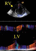
The AcuNav (TM)-catheter is now pressed against the lateral RV wall avoiding its motion within the right ventricular cavity. LV = left ventricle; RV = right ventricle Keywords: Catheter,
echocardiography,
Echokardiographie,
Katheter,
ventricle,
Ventrikel
|
|
|
11479. Bartel T;
Caspari G;
Erbel R;
Mueller S
Intracardiac Echocardiography - Technology and Clinical Role
Intrakardiale Echokardiographie
Journal of Clinical and Basic Cardiology 2002; 5 (2): 133-137
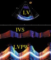
Rhythmic motion of the AcuNav (TM)-catheter within the right ventricle causes pseudoparadoxical motion of the interatrial septum. IVS = interventricular septum; LV = left ventricle; LVPW =
left ventricular posterior wall Keywords: Catheter,
echocardiography,
Echokardiographie,
Katheter,
Septum,
Septum,
ventricle,
Ventrikel
|
|
|
11480. Bartel T;
Caspari G;
Erbel R;
Mueller S
Intracardiac Echocardiography - Technology and Clinical Role
Intrakardiale Echokardiographie
Journal of Clinical and Basic Cardiology 2002; 5 (2): 133-137
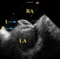
Device closure of a patent foramen ovale guided by
intracardiac echocardiography with the AcuNav )(TM)-catheter placed in the right atrium and retroflexed into a short-axis orientation. Typical attitude before the Amplatzer (TM)-PFO occluder is removed from the cable. LA = left atrium; RA = right atrium; 1, right umbrella of Amplatzer (TM)-PFO occluder; 2, left umbrella of Amplatzer (TM)-PFO occluder Keywords: Amplatzer-PFO-Occluder,
Amplatzer-PFO-Occluder,
Atrium,
Atrium,
Catheter,
echocardiography,
Echokardiographie,
Foramen ovale,
Foramen ovale,
Katheter
|
|
|
| |
|

 Suche Medizinische Publikationen
 Suche Abbildungen und Grafiken
 Medizinprodukte - Labortechnik
 Antidotarium (Stand 2012)
 Phytokodex (Stand 2012)
 Fachbücher
 Patientenliteratur