|
|
|---|
3241. Schweitzer N;
Vogel A
Medikamentöse Standardtherapie des Cholangiokarzinoms und aktuelle Arzneimittelstudien
Cholangiokarzinom
Journal für Gastroenterologische und Hepatologische Erkrankungen 2014; 12 (1): 11-17
Signalwege und Angriffspunkte der molekularen Therapien beim Cholangiokarzinom. Erstellt nach Daten aus [65]. EGRF: epidermal growth factor receptor; VEGFR: vascular endothelial growth factor receptor; RAS: rat sarcoma protein; PI3K: Phosphatidylinositol-3-Kinase; BRAF: v-Raf murine sarcoma viral oncogene homolog B1; PIP2: Phosphatidylinositol-4,5-Bisphosphat, PIP3: Phosphatidylinositol-3,4,5-Triphosphat; MEK: mitogen-activated protein kinase/extracellular-signal regulated kinase; PTEN: phosphatase and tensin homolog; Rheb: RAS homolog enriched in brain; TSC: tuberous sclerosis complex; AKT: protein kinase B; mTOR: mammalian target of rapamycin. Keywords: Cholangiokarzinom,
molekulare Therapie,
Schema
|
|
|
3242. Schneider N. I.;
Langner C
Histologie der Refluxkrankheit und ihre Bedeutung für die Entstehung des Barrett-Ösophagus
Barrett-Ösophagus
Journal für Gastroenterologische und Hepatologische Erkrankungen 2014; 12 (1): 5-10
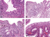
(a) Mehrschichtiges Epithel (multilayered epithelium) an der gastroösophagealen Grenze, charakterisiert durch basale Zellen mit plattenepithelialer Differenzierung und luminale
Zylinderepithelien (original ×200). (b, c) Kardiamukosa und mehrschichtiges Epithel (Pfeile)
im Ausführungsgang eines Drüsenläppchens;
Lokalisation der Biopsieentnahme somit im distalen
Ösophagus ([b] original ×100; [c]: Ausschnitt von [b] original ×200). (d) Mehrschichtiges Epithel (obere Bildhälfte) assoziiert mit klassischer becherzellhaltiger Barrett-Mukosa (untere Bildhälfte, original ×100). Keywords: Barrett-Ösophagus,
Histologie,
Refluxkrankheit
|
|
|
3243. Schneider N. I.;
Langner C
Histologie der Refluxkrankheit und ihre Bedeutung für die Entstehung des Barrett-Ösophagus
Refluxkrankheit
Journal für Gastroenterologische und Hepatologische Erkrankungen 2014; 12 (1): 5-10
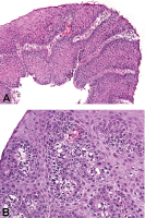
(a) Regeneratorische Veränderungen des Plattenepithels des Ösophagus bei chronischem Reflux (hyperregeneratorische Ösophagopathie) mit Verbreiterung der Basalzelllagen und Verlängerung der Stromapapillen (original ×100). (b) Dilatation der Interzellularspalten und leichte entzündliche Infiltration (original ×200). Keywords: Histologie,
Refluxkrankheit,
Ösophagus
|
|
|
3244. Traindl O;
Watschinger B
Kardiovaskuläre und renale Risikoreduktion durch lipidsenkende Therapie bei Patienten mit chronischer Niereninsuffizienz
Chronische Niereninsuffizienz
Journal für Hypertonie - Austrian Journal of Hypertension 2014; 18 (1): 9-17
Adjustierte Hazard Ratios für Tod und kardiovaskuläre Ereignisse in der Population US-amerikanischer Patienten mit chronischer Niereninsuffizienz. Mod. nach [10].
x-Achse: GFR entsprechend K/DOQI-CKD-Stadien [37], y-Achse: Hazard Ratio. Keywords: Diagramm,
Hazard Ratio,
Hypertonie,
Niereninsuffizienz
|
|
|
3245. Brezinka C
Tipps und Tricks im Gyn-Ultraschall: Wie sicher ist die Abortus-Diagnostik im Ultraschall? Ab wann darf man eine Schwangerschaft „aufgeben“?
Gyn-Ultraschall
Journal für Gynäkologische Endokrinologie 2014; 8 (1) (Ausgabe für Österreich): 24-27
Journal für Gynäkologische Endokrinologie 2014; 8 (1) (Ausgabe für Schweiz): 26-29
(a) Ein 3 mm großer Embryo ohne Herzaktion; (b) 2 Tage später: Das HCG ist gesunken, der Fruchtsack ist geschrumpft. Keywords: Abortusdiagnose,
Gynäkologie,
Sonographie,
Ultraschall
|
|
|
3246. Brezinka C
Tipps und Tricks im Gyn-Ultraschall: Wie sicher ist die Abortus-Diagnostik im Ultraschall? Ab wann darf man eine Schwangerschaft „aufgeben“?
Gyn-Ultraschall
Journal für Gynäkologische Endokrinologie 2014; 8 (1) (Ausgabe für Österreich): 24-27
Journal für Gynäkologische Endokrinologie 2014; 8 (1) (Ausgabe für Schweiz): 26-29
Ein 9 mm großer Embryo ohne Herzaktion. Keywords: Abortusdiagnose,
Gynäkologie,
Sonographie,
Ultraschall
|
|
|
3247. Brezinka C
Tipps und Tricks im Gyn-Ultraschall: Wie sicher ist die Abortus-Diagnostik im Ultraschall? Ab wann darf man eine Schwangerschaft „aufgeben“?
Gyn-Ultraschall
Journal für Gynäkologische Endokrinologie 2014; 8 (1) (Ausgabe für Österreich): 24-27
Journal für Gynäkologische Endokrinologie 2014; 8 (1) (Ausgabe für Schweiz): 26-29
Ein im Mittel 24 mm großer Fruchtsack in der SSW 8+4 ohne Herzaktion und mit sinkendem HCG: Befund einer „missed abortion“. Keywords: Abortusdiagnose,
Gynäkologie,
Sonographie,
Ultraschall
|
|
|
3248. Brezinka C
Tipps und Tricks im Gyn-Ultraschall: Wie sicher ist die Abortus-Diagnostik im Ultraschall? Ab wann darf man eine Schwangerschaft „aufgeben“?
Gyn-Ultraschall
Journal für Gynäkologische Endokrinologie 2014; 8 (1) (Ausgabe für Österreich): 24-27
Journal für Gynäkologische Endokrinologie 2014; 8 (1) (Ausgabe für Schweiz): 26-29
Ein 14 mm großer Fruchtsack, keine embryonalen Anteile, kein Dottersack. Die rundliche Struktur rechts im Bild ist das Corpus luteum. Keywords: Abortusdiagnose,
Gynäkologie,
Sonographie,
Ultraschall
|
|
|
3249. Brezinka C
Tipps und Tricks im Gyn-Ultraschall: Wie sicher ist die Abortus-Diagnostik im Ultraschall? Ab wann darf man eine Schwangerschaft „aufgeben“?
Gyn-Ultraschall
Journal für Gynäkologische Endokrinologie 2014; 8 (1) (Ausgabe für Österreich): 24-27
Journal für Gynäkologische Endokrinologie 2014; 8 (1) (Ausgabe für Schweiz): 26-29
(a) Abortgeschehen in der 8. SSW mit 3 cm Hämatom/Koageln im Cavum uteri ohne erkennbaren Fruchtsack; (b) Darstellung mit der Abdominalsonde. Keywords: Abortusdiagnose,
Gynäkologie,
Sonographie,
Ultraschall
|
|
|
3250. Brezinka C
Tipps und Tricks im Gyn-Ultraschall: Wie sicher ist die Abortus-Diagnostik im Ultraschall? Ab wann darf man eine Schwangerschaft „aufgeben“?
Gyn-Ultraschall
Journal für Gynäkologische Endokrinologie 2014; 8 (1) (Ausgabe für Österreich): 24-27
Journal für Gynäkologische Endokrinologie 2014; 8 (1) (Ausgabe für Schweiz): 26-29
Ein 4 mm großer topischer Fruchtsack am 5. Tag der Amenorrhö im linken Anteil eines leichten Uterus arcuatus. So früh kann man noch keine Diagnose und keine Aussagen über die Prognose der Schwangerschaft machen. Keywords: Abortusdiagnose,
Gynäkologie,
Sonographie,
Ultraschall
|
|
|
3251. Moser W;
Marhold F;
Ungersböck K;
Oberndorfer S
Subacute Spinal Cord Cempression Due to an Intramedullar Spinal Teratoma in a 53-Year-Old Female Patient: A Case Report
Spinal Teratoma
European Association of NeuroOncology Magazine 2014; 4 (1): 34-36
MRI of the thoracic and lumbar spine after partial resection. Keywords: MRI,
spinal teratoma
|
|
|
3252. Moser W;
Marhold F;
Ungersböck K;
Oberndorfer S
Subacute Spinal Cord Cempression Due to an Intramedullar Spinal Teratoma in a 53-Year-Old Female Patient: A Case Report
Spinal Teratoma
European Association of NeuroOncology Magazine 2014; 4 (1): 34-36
Intraoperative pictures: (a) after opening of the dura mater; (b) pus-like liquid appearing during
resection of the tumour; (c) collapse of the myelon
after resection. Keywords: intraoperative picture,
photograph,
spinal teratoma
|
|
|
3253. Moser W;
Marhold F;
Ungersböck K;
Oberndorfer S
Subacute Spinal Cord Cempression Due to an Intramedullar Spinal Teratoma in a 53-Year-Old Female Patient: A Case Report
Spinal Teratoma
European Association of NeuroOncology Magazine 2014; 4 (1): 34-36
MRI of the thoracic and lumbar spine at admission, showing the teratoma at levels Th11/Th12. Keywords: MRI,
spinal teratoma
|
|
|
3254. Grossman R;
Ram Z
Awake Craniotomy in Glioma Surgery
Awake Craniotomy
European Association of NeuroOncology Magazine 2014; 4 (1): 27-33
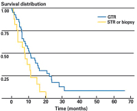
Kaplan-Meier curve of survival for elderly patients (65 years) with highgrade gliomas (HGG) who underwent awake-craniotomy gross tumour resection (GTR) compared to subtotal resection (STR) or biopsy. Median survival was significantly longer for patients with HGG who underwent GTR compared to those who underwent STR or biopsy (10.8 vs 7.8 months, respectively; p = 0.039) [39]. Keywords: awake craniotomy,
diagram,
glioma surgery,
Kaplan-Meier,
survival distribution
|
|
|
3255. Grossman R;
Ram Z
Awake Craniotomy in Glioma Surgery
Awake Craniotomy
European Association of NeuroOncology Magazine 2014; 4 (1): 27-33
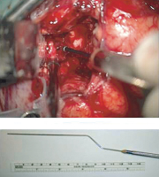
Sub-cortical stimulation of the cortico-spinal tracts by means of a subcortical monopolar electrode. Keywords: awake craniotomy,
cortical stimulation,
glioma surgery,
photograph
|
|
|
3256. Grossman R;
Ram Z
Awake Craniotomy in Glioma Surgery
Awake Craniotomy
European Association of NeuroOncology Magazine 2014; 4 (1): 27-33
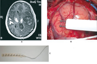
(A) Preoperative T1-weighted axial after gadolinium injection with diffuse tensor imaging (DTI) of the cortico-spinal tract showing a left temporo-parietal high-grade glioma displace medially the corticospinal tract.
(B) Direct cortical stimulation by using bipolar probe.
(C) A cortical strip electrode is placed over the surface of the motor cortex in order to assess motor-evoked potential (MEP). Keywords: awake craniotomy,
cortical stimulation,
glioma surgery,
MRI,
photograph
|
|
|
3257. Grossman R;
Ram Z
Awake Craniotomy in Glioma Surgery
Awake Craniotomy
European Association of NeuroOncology Magazine 2014; 4 (1): 27-33
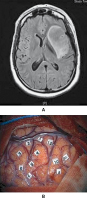
(A) Pre-operative axial fluid-attenuated inversion recovery- (FLAIR-) weighted MRI showing a WHO grade-II insular glioma in a 40-year-old woman presenting with episodes of seizures. This patient has no neurological deficit. (B) Intra-operative
cortical mapping of language during awake surgery. Keywords: awake craniotomy,
cortical mapping,
MRI,
photograph
|
|
|
3258. Grossman R;
Ram Z
Awake Craniotomy in Glioma Surgery
Awake Craniotomy
European Association of NeuroOncology Magazine 2014; 4 (1): 27-33
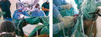
Intra-operative photographs of the surgical team during awake craniotomy. Keywords: awake craniotomy,
glioma surgery,
photograph
|
|
|
3259. Uckermann O;
Galli R;
Mackenroth L;
Geiger K;
Steiner G;
Koch E;
Schackert G;
Kirsch M
Optical Biochemical Imaging: Potential New Applications in Neuro-Oncology
NLO imaging
European Association of NeuroOncology Magazine 2014; 4 (1): 20-26
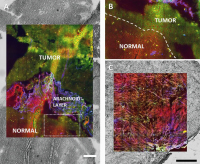
Multimodal NLO imaging of human tumours (red: CARS, green: TPEF, blue: SHG), overlaid with the bright field images of the unstained sample. The technique allows to retrieve detailed morphochemical information about tissue structure and properties on unstained samples. (a) Cryosection of human glioblastoma (scale bar: 200 μm). (b) High magnification of the border between tumour and normal tissue as indicated in (a). (c) Cryosection of human neuroma (scale bar: 0.5 mm). Keywords: NLO imaging,
nonlinear optical microscopy
|
|
|
3260. Uckermann O;
Galli R;
Mackenroth L;
Geiger K;
Steiner G;
Koch E;
Schackert G;
Kirsch M
Optical Biochemical Imaging: Potential New Applications in Neuro-Oncology
Multimodal NLO
European Association of NeuroOncology Magazine 2014; 4 (1): 20-26
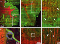
Multimodal NLO imaging of a mouse cerebellum (red: CARS, green: TPEF, blue: SHG). (a) Overview of the mouse cerebellum. (b) Magnification of the area indicated in (a). (c) Magnification of the area indicated in (b). Arrows indicate Purkinje cells characterized by punctuate fluorescence in the somata. (d) Magnification of
the area indicated in (a), a large blood vessel is shown (*). (e) Magnification of the area indicated in (a), nerve fibres are indicated by intense CARS signal. (f) Magnification of the area indicated in (e). Single axons can be discerned (arrows). Keywords: NLO imaging,
nonlinear optical microscopy
|
|
|
3261. Uckermann O;
Galli R;
Mackenroth L;
Geiger K;
Steiner G;
Koch E;
Schackert G;
Kirsch M
Optical Biochemical Imaging: Potential New Applications in Neuro-Oncology
CARS image
European Association of NeuroOncology Magazine 2014; 4 (1): 20-26
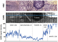
CARS microscopy of infiltrative experimental
glioblastoma in a mouse model: The intensity of the CARS image and the morphological details permit to distinguish between the tumour mass, the infiltrative area, and the normal brain grey and white matters. H&E staining (top) CARS image (centre). The intensity of the CARS signal along the blue line is plotted in the graph (bottom). Mean intensity values for the different types of tissue are also reported. Mean intensity that characterizes the normal grey tissue is reduced to approximately 50 % in the infiltrative area and to approximately 30 % in the tumour. Lipid rich white matter is characterized by high CARS signal intensity (228, saturation in some areas). Keywords: CARS image,
scheme
|
|
|
3262. Uckermann O;
Galli R;
Mackenroth L;
Geiger K;
Steiner G;
Koch E;
Schackert G;
Kirsch M
Optical Biochemical Imaging: Potential New Applications in Neuro-Oncology
FT-IR imaging
European Association of NeuroOncology Magazine 2014; 4 (1): 20-26
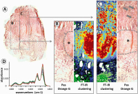
FT-IR imaging of human pituitary adenoma. (a) Overview of a cryosection of human pituitary adenoma stained using Pas-Orange-G and illustrating the production of human growth hormone (HGH). (b, c) High magnification of the areas indicated in (a) and corresponding FT-IR spectroscopic images obtained by cluster analysis. Large areas with increased HGH production are indicated by asterisks, local spots of hormone production are marked by arrows. (d) Representative spectra corresponding to area (b). Keywords: FT-IR imaging,
human pituitary adenoma,
scheme
|
|
|
3263. Uckermann O;
Galli R;
Mackenroth L;
Geiger K;
Steiner G;
Koch E;
Schackert G;
Kirsch M
Optical Biochemical Imaging: Potential New Applications in Neuro-Oncology
Vibrational Spectroscopy
European Association of NeuroOncology Magazine 2014; 4 (1): 20-26
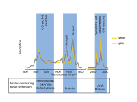
FT-IR spectra of grey and white matters of nervous tissue. The main bond vibrations and related dominating tissue components are indicated. Keywords: FT-IR spectra,
scheme,
vibrational spectroscopy
|
|
|
3264. Schwabl C;
Gaugg M;
Beham-Schmid C;
Celedin S;
Liegl-Atzwanger B;
Matschnig S;
Raunik W;
Weiss H;
Neumann HJ
Rheumatoide Arthritis und seltene Tumormanifestation – auf Umwegen zur Remission
Follikuläres Lymphom
Tumorboard 2014; 3 (1): 5-9
Relativ niedrige Proliferationsfraktion mit typischem Verteilungsmuster. MIB1 x 600 Keywords: Biopsie,
follikuläres Lymphom,
Onkologie,
Proliferationsfraktion
|
|
|
3265. Schwabl C;
Gaugg M;
Beham-Schmid C;
Celedin S;
Liegl-Atzwanger B;
Matschnig S;
Raunik W;
Weiss H;
Neumann HJ
Rheumatoide Arthritis und seltene Tumormanifestation – auf Umwegen zur Remission
Follikuläres Lymphom
Tumorboard 2014; 3 (1): 5-9
Starke Reaktivität der infiltrierenden Zellen mit CD20. x 600. Keywords: Biopsie,
follikuläres Lymphom,
Immunhistochemie,
Onkologie
|
|
|
3266. Schwabl C;
Gaugg M;
Beham-Schmid C;
Celedin S;
Liegl-Atzwanger B;
Matschnig S;
Raunik W;
Weiss H;
Neumann HJ
Rheumatoide Arthritis und seltene Tumormanifestation – auf Umwegen zur Remission
Follikuläres Lymphom
Tumorboard 2014; 3 (1): 5-9
Bei starker Vergrößerung besteht das lymphatische Infiltrat aus kleinen Lymphozyten mit gebuchteten und gekerbten Kernen sowie untermischten Blasten. HE, x 600 Keywords: Biopsie,
follikuläres Lymphom,
lymphatisches Infiltrat,
Onkologie
|
|
|
3267. Schwabl C;
Gaugg M;
Beham-Schmid C;
Celedin S;
Liegl-Atzwanger B;
Matschnig S;
Raunik W;
Weiss H;
Neumann HJ
Rheumatoide Arthritis und seltene Tumormanifestation – auf Umwegen zur Remission
Follikuläres Lymphom
Tumorboard 2014; 3 (1): 5-9
Biopsie (aus M. iliacus) mit Sklerosierung und dichter lymphatischer Infiltration. HE-Färbung, x 25. Keywords: Biopsie,
follikuläres Lymphom,
lymphatische Infiltration,
Onkologie
|
|
|
3268. Oberndorfer S
Tumorboards stellen sich vor: Neuroonkologisches Tumorboard
Neuroonkologische Tumorboard
Tumorboard 2014; 3 (1): 21-23
Zusammensetzung eines neuroonkologischen Tumorboards. Keywords: neuroonkologisches Tumorboard,
Onkologie,
Schema,
Tumorboard
|
|
|
3269. Mayer-Pickel K;
Benedicic C;
Lang U;
Tamussino K;
Petru E
Metastasiertes Magenkarzinom in der Schwangerschaft – ein Fallbericht
Magenkarzinom
Tumorboard 2014; 3 (1): 19-20
Niedrig differenziertes Adenokarzinom des Magens Keywords: Adenokarzinom,
Histologie,
Magenkarzinom,
Onkologie
|
|
|
3270. Mayer-Pickel K;
Benedicic C;
Lang U;
Tamussino K;
Petru E
Metastasiertes Magenkarzinom in der Schwangerschaft – ein Fallbericht
Krukenberg-Tumor
Tumorboard 2014; 3 (1): 19-20
Krukenberg-Tumor des rechten Ovars, ausgehend von einem niedrig differenzierten Adenokarzinom Keywords: Adenokarzinom,
Histologie,
Krukenberg-Tumor,
Onkologie
|
|
|
3271. Mayer-Pickel K;
Benedicic C;
Lang U;
Tamussino K;
Petru E
Metastasiertes Magenkarzinom in der Schwangerschaft – ein Fallbericht
Magenkarzinom
Tumorboard 2014; 3 (1): 19-20
Großer expansiver Prozess im rechten Unterbauch in der 20. Schwangerschaftswoche Keywords: expansiver Prozess,
MR,
Onkologie,
Unterbauch
|
|
|
3272. Schmied B;
Reitsamer R;
Gschwandtner E;
Farmini A;
Fischer T;
Kronberger C;
Peintinger F
Metaplastisches Mammakarzinom – ein Fallbericht und Review der aktuellen Literatur
Mammakarzinom
Tumorboard 2014; 3 (1): 16-18
Liegende Bülau-Drainage und liegende Pigtail-Drainage – Verlaufsröntgen bei zunehmender respiratorischer Insuffizienz Keywords: Drainage,
Mammakarzinom,
Onkologie,
Röntgen
|
|
|
3273. Schmied B;
Reitsamer R;
Gschwandtner E;
Farmini A;
Fischer T;
Kronberger C;
Peintinger F
Metaplastisches Mammakarzinom – ein Fallbericht und Review der aktuellen Literatur
Mammakarzinom
Tumorboard 2014; 3 (1): 16-18
Lokalbefund nach R2-Resektion Keywords: Foto,
Mammakarzinom,
Onkologie,
Resektion
|
|
|
3274. Schmied B;
Reitsamer R;
Gschwandtner E;
Farmini A;
Fischer T;
Kronberger C;
Peintinger F
Metaplastisches Mammakarzinom – ein Fallbericht und Review der aktuellen Literatur
Mammakarzinom
Tumorboard 2014; 3 (1): 16-18
Fotodokumentation nach 4 Zyklen Chemotherapie (3x Epirubicin/Docetaxel und 1x Carboplatin/Gemcitabin) Keywords: Chemotherapie,
Foto,
Mammakarzinom,
Onkologie
|
|
|
3275. Schmied B;
Reitsamer R;
Gschwandtner E;
Farmini A;
Fischer T;
Kronberger C;
Peintinger F
Metaplastisches Mammakarzinom – ein Fallbericht und Review der aktuellen Literatur
Mammakarzinom
Tumorboard 2014; 3 (1): 16-18
Mamma-MRT nach 2 Zyklen Chemotherapie Keywords: Chemotherapie,
Mammakarzinom,
MRT,
Onkologie
|
|
|
3276. Schmied B;
Reitsamer R;
Gschwandtner E;
Farmini A;
Fischer T;
Kronberger C;
Peintinger F
Metaplastisches Mammakarzinom – ein Fallbericht und Review der aktuellen Literatur
Mammakarzinom
Tumorboard 2014; 3 (1): 16-18
Stanzbiopsie: Links: Infiltration des metaplastischen Tumors (*) in die Muskulatur (o) (Vergrößerung 4x). Rechts: Detailansicht des mitotisch hoch aktiven gering differenzierten Tumoranteils (Vergrößerung 25x) Keywords: Mammakarzinom,
metaplastischer Tumor,
Onkologie,
Stanzbiopsie
|
|
|
3277. Schmied B;
Reitsamer R;
Gschwandtner E;
Farmini A;
Fischer T;
Kronberger C;
Peintinger F
Metaplastisches Mammakarzinom – ein Fallbericht und Review der aktuellen Literatur
Mammakarzinom
Tumorboard 2014; 3 (1): 16-18
Fotodokumentation bei Erstvorstellung Keywords: Brustwandinfiltration,
Foto,
Mammakarzinom,
Onkologie,
palpabler Tumor
|
|
|
3278. Mayer-Pickel K;
Benedicic C;
Pristauz G;
Lang U;
Tamussino K;
Petru E
Metastasiertes Mammakarzinom in der Schwangerschaft mit fulminantem Verlauf – Ein Fallbericht
Wirbelkörpermetastasen
Tumorboard 2014; 3 (1): 13-15
Wirbelkörpermetastase BWK-10, 11, 12 und LWK-4, dargestellt in der seitlichen Röntgenaufnahme Keywords: MR,
Onkologie,
Röntgen,
Wirbelkörpermetastasen
|
|
|
3279. Mayer-Pickel K;
Benedicic C;
Pristauz G;
Lang U;
Tamussino K;
Petru E
Metastasiertes Mammakarzinom in der Schwangerschaft mit fulminantem Verlauf – Ein Fallbericht
Thoraxröntgen
Tumorboard 2014; 3 (1): 13-15
Basaler Pleuraerguß rechts Keywords: Onkologie,
Pleuraerguss,
Röntgen,
Thorax
|
|
|
3280. Mayer-Pickel K;
Benedicic C;
Pristauz G;
Lang U;
Tamussino K;
Petru E
Metastasiertes Mammakarzinom in der Schwangerschaft mit fulminantem Verlauf – Ein Fallbericht
Mammakarzinom
Tumorboard 2014; 3 (1): 13-15
Suspekter Knoten der linken Brust in der Mammasonographie Keywords: Mammasonographie,
Onkologie,
suspekter Knoten
|
|
|
| |

 Suche Medizinische Publikationen
 Suche Abbildungen und Grafiken
 Medizinprodukte - Labortechnik
 Antidotarium (Stand 2012)
 Phytokodex (Stand 2012)
 Fachbücher
 Patientenliteratur