|
|
|---|
1. Zweiker D;
Santner V;
Nürnberg M;
Scherr D
Die Defibrillatorweste im Laufe der Jahre // The wearable cardioverter defibrillator over the years
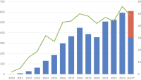
WCD-Erstverschreibungen
Journal für Kardiologie - Austrian Journal of Cardiology 2025; 32 (5-6): 92-96
WCD-Erstverschreibungen pro Jahr (blau, primäre Achse) und Anzahl der verschreibenden Zentren (grün, sekundäre Achse). *Die Anzahl der Verschreibungen im Jahr 2024 (Daten bis Juli 2024 verfügbar) wurde auf 12 Monate extrapoliert (orange). Keywords: Defibrillatorweste,
Diagramm,
Kardiologie,
Verschreibung,
WCD
|
|
|
2. Manninger-Wünscher M;
Geczy T;
Scherr D
EPU-Corner: Elektroporation zur Pulmonalvenenisolation bei Vorhofflimmern
Elektroanatomisches Mapping
Journal für Kardiologie - Austrian Journal of Cardiology 2021; 28 (11-12): 406-407
Elektroanatomisches Mapping vor (oben) und nach
(unten) Ablation. Links: Ansicht von anterior und rechts lateral;
rechts: Ansicht von posterior und links lateral. Farbskalierung:
violett = normale Voltage, rot < 0,2 mV. Keywords: Elektroanatomisches Mapping,
Kardiologie
|
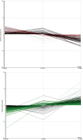
|
|
3. Manninger-Wünscher M;
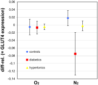
Geczy T;
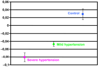
Scherr D
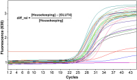
EPU-Corner: Elektroporation zur Pulmonalvenenisolation bei Vorhofflimmern
Fluoroskopie
Journal für Kardiologie - Austrian Journal of Cardiology 2021; 28 (11-12): 406-407
Fluoroskopie-Bilder der Ablation der linken unteren
Pulmonalvene in „basket“- (links, AP-Projektion) und „flower“-
Konfiguration (rechts, LAO 60° Projektion). Zwei Diagnostikkatheter im rechten Ventrikel und Koronarsinus.
Keywords: Fluoroskopie,
Kardiologie
|
|
|
4. Manninger-Wünscher M;
Geczy T;
Scherr D
EPU-Corner: Elektroporation zur Pulmonalvenenisolation bei Vorhofflimmern
Kontrastmittel-CT
Journal für Kardiologie - Austrian Journal of Cardiology 2021; 28 (11-12): 406-407
Das segmentierte Kontrastmittel-CT des linken
Atriums zeigt eine normale Anatomie mit jeweils zwei linken und
rechten Pulmonalvenen. Links: Ansicht von posterior; rechts: Ansicht von links lateral Keywords: Kardiologie,
Kontrastmittel-CT
|
|
|
5. Zweiker D;
Manninger M;
Scherr D
Telemedizin bei „Wearables“ // Remote care in wearables
Vorhofflimmern
Journal für Kardiologie - Austrian Journal of Cardiology 2021; 28 (11-12): 386-389
6-Kanal-Wearable-EKG-Aufzeichnung einer Arrhythmie. In der Aufzeichnung zeigt sich eine arrhythmische Schmalkomplextachykardie mit einer Frequenz von 170/min. Die Aufzeichnung ist diagnostisch für Vorhofflimmern. Keywords: Kardiologie,
Schmalkomplextachykardie,
Vorhofflimmern
|
|
|
6. Zweiker D;
Manninger M;
Scherr D
Telemedizin bei „Wearables“ // Remote care in wearables
Vorhofflimmerarrhythmie
Journal für Kardiologie - Austrian Journal of Cardiology 2021; 28 (11-12): 386-389
Die Aufzeichnung für 2 Minuten zeigt eine unregelmäßige Pulskurve (Bild oben) sowie deutlich unregelmäßige RR-Abstände (Bild links unten) und eine große Streuung im Poincaré-Plot (Bild rechts unten). In Zusammenschau der Befunde besteht der
Verdacht auf eine Vorhofflimmerarrhythmie. Keywords: Kardiologie,
Poincaré-Plot,
Vorhofflimmerarrhythmie
|
|
|
7. Manninger-Wünscher M;
Bisping E;
Lercher P;
Prenner G;
Rohrer U;
Sereinigg M;
Zirlik A;
Scherr D
EPU-Corner: Elektrischer Sturm bei WPW-Syndrom
AAI-Stimulation
Journal für Kardiologie - Austrian Journal of Cardiology 2020; 27 (7-8): 302-305
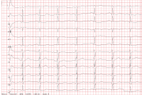
12-Kanal-EKGs unter laufender AAI-Stimulation vor (A) und nach (B) Ablation der beiden akzessorischen Bahnen.
Keywords: AAI-Stimulation,
EKG,
Kardiologie
|
|
|
8. Manninger-Wünscher M;
Bisping E;
Lercher P;
Prenner G;
Rohrer U;
Sereinigg M;
Zirlik A;
Scherr D
EPU-Corner: Elektrischer Sturm bei WPW-Syndrom
Ablation
Journal für Kardiologie - Austrian Journal of Cardiology 2020; 27 (7-8): 302-305
Ablation der links lateralen Bahn führt zu Verlust der Delta-Welle im Oberflächen-EKG (hier V3 und V4) 5 Sekunden nach Start der Ablation. Keywords: Ablation,
EKG,
Kardiologie
|
|
|
9. Manninger-Wünscher M;
Bisping E;
Lercher P;
Prenner G;
Rohrer U;
Sereinigg M;
Zirlik A;
Scherr D
EPU-Corner: Elektrischer Sturm bei WPW-Syndrom
Katheterablation
Journal für Kardiologie - Austrian Journal of Cardiology 2020; 27 (7-8): 302-305
Katheterablation der posteroseptalen (A: RAO, B: LAO) und links lateralen Bahn (C: RAO, D: LAO). Rot: Ablationskatheter; blau: Diagnostikkathe ter im hohen rechten Atrium; gelb: His-Katheter; violett: Diagnostikkatheter im rechten Ventrikel; grün: Diagnostikkatheter im Koronarsinus.
Keywords: Echo,
Kardiologie,
Katheterablation
|
|
|
10. Manninger-Wünscher M;
Bisping E;
Lercher P;
Prenner G;
Rohrer U;
Sereinigg M;
Zirlik A;
Scherr D
EPU-Corner: Elektrischer Sturm bei WPW-Syndrom
FBI-Tachykardie
Journal für Kardiologie - Austrian Journal of Cardiology 2020; 27 (7-8): 302-305
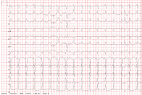
12-Kanal-EKG der FBI-Tachykardie mit minimalen RR-Abständen von bis zu 200 ms. Keywords: EKG,
FBI-Tachykardie,
Kardiologie
|
|
|
11. Scherr D;
Manninger-Wünscher M
COVID-19 und Arrhythmien // COVID-19 and arrhythmias
Therapiemanagement
Journal für Kardiologie - Austrian Journal of Cardiology 2020; 27 (5): 168-170
Management bei der Therapie von COVID-19 mit QTc-verlängernden Medikamenten. Erstellt nach der ESC-COVID-Richtlinie auf www.escardio.org. © D. ScherrKeywords: Arrhythmie,
COVID-19,
Kardiologie,
Management,
QTc-Verlängerung,
Schema,
Therapie
|
|
|
12. Manninger-Wünscher M;
Rohrer U;
Toller W;
Maier A;
Theuermann C;
Bisping E;
Lercher P;
Prenner G;
Sereinigg M;
Smolle-Jüttner FM;
Zirlik A;
Scherr D
EPU-Corner: Sympathektomie als Bailout-Therapie bei elektrischem Sturm
Sympathektomie
Journal für Kardiologie - Austrian Journal of Cardiology 2019; 26 (11-12): 332-333
Sympathektomie. Links: thorakoskopischer Zugang; rechts: Darstellung des N. sympathicus vor segmentaler Resektion
(roter Pfeil) Keywords: Kardiologie,
Sympathektomie
|
|
|
13. Manninger-Wünscher M;
Rohrer U;
Toller W;
Maier A;
Theuermann C;
Bisping E;
Lercher P;
Prenner G;
Sereinigg M;
Smolle-Jüttner FM;
Zirlik A;
Scherr D
EPU-Corner: Sympathektomie als Bailout-Therapie bei elektrischem Sturm
Epikardiales Mapping
Journal für Kardiologie - Austrian Journal of Cardiology 2019; 26 (11-12): 332-333
Epikardiales Mapping des linken Ventrikels. Links: modifizierte Ansicht in links lateral; rechts: modifizierte Ansicht in LAO.
Normale Voltage zeigt sich in violett, Narbenareale in rot (Voltage-Cutoff 0,05 mV). Basolateral wurde eine Ablationslinie gesetzt. Keywords: Epikardiales Mapping,
Kardiologie
|
|
|
14. Manninger-Wünscher M;
Rohrer U;
Toller W;
Maier A;
Theuermann C;
Bisping E;
Lercher P;
Prenner G;
Sereinigg M;
Smolle-Jüttner FM;
Zirlik A;
Scherr D
EPU-Corner: Sympathektomie als Bailout-Therapie bei elektrischem Sturm
EKG
Journal für Kardiologie - Austrian Journal of Cardiology 2019; 26 (11-12): 332-333
12-Kanal-EKG der Kammertachykardie. Das EKG
zeigt eine inferiore Achse (positiv in II, III, aVF) sowie eine Transition
in V3. Keywords: EKG,
Kardiologie
|
|
|
15. Zweiker D;
Bisping E;
Lercher P;
Manninger M;
Prenner G;
Riedlbauer R;
Scherr D
EPU-Corner: Incessant WPW-Syndrom
Elektrophysiologische Untersuchung
Journal für Kardiologie - Austrian Journal of Cardiology 2018; 25 (5-6): 148-149
Elektrophysiologische Untersuchung. Es zeigt sich unter Adenosin-Gabe (12 mg i. v.) und rechtsventrikulärer Stimulation eine retrograde ventrikuloatriale Überleitung. Das erste atriale Signal ist bei CS 3/4 sichtbar. Keywords: Adenosin-Gabe,
Elektrophysiologische Untersuchung,
Kardiologie,
rechtsventrikuläre Stimulation,
WPW-Syndrom
|
|
|
16. Zweiker D;
Bisping E;
Lercher P;
Manninger M;
Prenner G;
Riedlbauer R;
Scherr D
EPU-Corner: Incessant WPW-Syndrom
EKG nach Kardioversion
Journal für Kardiologie - Austrian Journal of Cardiology 2018; 25 (5-6): 148-149
Elektrokardiogramm nach elektrischer Kardioversion. Sinusrhythmus, Frequenz 90/min, Steiltyp, PQ-Dauer 140 ms, QRS-Dauer 80 ms, R/S-Umschlag in V5, normale Repolarisation, QT 390ms, QTc 476ms. Keywords: EKG,
Elektrokardiogramm,
Kardiologie,
Kardioversion,
WPW-Syndrom
|
|
|
17. Zweiker D;
Bisping E;
Lercher P;
Manninger M;
Prenner G;
Riedlbauer R;
Scherr D
EPU-Corner: Incessant WPW-Syndrom
EKG bei Aufnahme
Journal für Kardiologie - Austrian Journal of Cardiology 2018; 25 (5-6): 148-149
Elektrokardiogramm bei der Aufnahme auf die Intensivstation. Rhythmische Schmalkomplextachykardie, Frequenz 198/min, Rechtstyp, Fusion der P- und T-Wellen, QRS-Dauer 70 ms, R/S-Umschlag in V5, negative Ts in II, III, aVF, QT 270ms, QTc 402ms. Keywords: Aufnahme,
EKG,
Elektrokardiogramm,
Kardiologie,
WPW-Syndrom
|
|
|
18. Manninger M;
Scherr D
EPU-Corner: Linksatriale Tachykardie nach Vorhofflimmerablation
Atriale Tachykardie
Journal für Kardiologie - Austrian Journal of Cardiology 2016; 23 (5-6): 149-150
Terminierung der atrialen Tachykardie während lokaler Ablation am
Vorhofdach am Oberrand des linken Herzohrs. (Schreibgeschwindigkeit 50 mm/s). Keywords: atriale Tachykardie,
Kardiologie
|
|
|
19. Manninger M;
Scherr D
EPU-Corner: Linksatriale Tachykardie nach Vorhofflimmerablation
Mikro-Reentry
Journal für Kardiologie - Austrian Journal of Cardiology 2016; 23 (5-6): 149-150
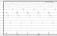
Nachweis des Mikro-Reentrys am Lasso-Katheter während der Erstellung
des Aktivierungsmaps. Der Lasso-Pol 6/7 befindet sich an der Basis des Vorhofdachs
(Punkt der frühesten Erregung der AT) und zeigt ein fraktioniertes Signal über
>100 ms als Ausdruck eines lokalen Mikro-Reentrys. Kanäle von oben nach unten:
Oberflächen-EKG II, bipolare Ableitungen über Lasso-Katheterspitze (1–2) bis proximal
(9–10), Ablationskatheter proximal, Referenz im Koronarsinus proximal (9–10).
(Schreibgeschwindigkeit 200 mm/s). Keywords: Aktivierungsmap,
Kardiologie,
Mikro-Reentry
|
|
|
20. Manninger M;
Scherr D
EPU-Corner: Linksatriale Tachykardie nach Vorhofflimmerablation
Aktivierungsmap
Journal für Kardiologie - Austrian Journal of Cardiology 2016; 23 (5-6): 149-150
CARTO® 3D-Aktivierungsmap (farbcodierte Darstellung der Aktivierungssequenz
im linken Atrium) der atrialen Tachykardie aufgenommen innerhalb von
nur 3 Minuten mittels Multielektrodenmapping über Lasso-Katheter: Nachweis der
frühesten Erregung am linken Vorhofdach am Herzohroberrand. Das lokale Signal an
diesem Punkt ist im Detail in Abbildung 3 dargestellt (Lasso-Pol 6/7).
(Ansicht des linken Atriums in ap; LAA = linkes Herzohr; LSPV = linke obere Pulmonalvene;
RSPV, RIPV = rechte obere bzw. untere Pulmonalvene, früheste Erregung = rot). Keywords: Aktivierungsmap,
atriale Tachykardie,
Kardiologie
|
|
|
21. Manninger M;
Scherr D
EPU-Corner: Linksatriale Tachykardie nach Vorhofflimmerablation
Atriale Tachykardie
Journal für Kardiologie - Austrian Journal of Cardiology 2016; 23 (5-6): 149-150
Atriale Tachykardie vor Ablation: Tachykardiezykluslänge von 422 ms,
fraktioniertes Signal mit einer Vorzeitigkeit von 92 ms am distalen Pol des Ablationskatheters
(ABL d). Kanäle von oben nach unten: Oberflächen-EKG I, II, III, V1, Ablationskatheter
distal, Koronarsinuskatheter von distal (CS 1,2) nach proximal (CS 9,10).
(Schreibgeschwindigkeit 100 mm/s). Keywords: Ablation,
atriale Tachykardie,
Kardiologie
|
|
|
22. Gasser R;
Ablasser K;
Roessl U;
Kraigher-Krainer E;
Lewinski D;
Mangge H;
Mächler H;
Trantina-Yates A;
Tscheliessnigg KH;
Udermann H;
Porta S;
Friehs I;
Scherr E;
Brussee H;
Gasser S
Differential Gene Expression und Nebivolol and Atenolol during Experimental Ischemia in Human Myocardium
Nebivolol - Biological Pathway
Journal of Clinical and Basic Cardiology 2008; 11 (1-4): 16-23
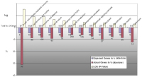
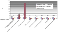
Concerning nebivolol-regulated biological pathways, we could see that, during normoxia, up-regulation of pathways is hardly pronounced by nebivolol over atenolol. This becomes evident also in Figure 9a, only 3
pathways are affected here. Noteworthy here, too, the interleukin signalling pathway. Keywords: Atenolol,
flowchart,
Nebivolol
|
|
|
23. Gasser R;
Ablasser K;
Roessl U;
Kraigher-Krainer E;
Lewinski D;
Mangge H;
Mächler H;
Trantina-Yates A;
Tscheliessnigg KH;
Udermann H;
Porta S;
Friehs I;
Scherr E;
Brussee H;
Gasser S
Differential Gene Expression und Nebivolol and Atenolol during Experimental Ischemia in Human Myocardium
Pathway - Up-Regulation
Journal of Clinical and Basic Cardiology 2008; 11 (1-4): 16-23
Generally, we could see that, during experimental ischemia, upregulation of pathways is minimal by nebivolol over atenolol. This becomes
evident in Figure 9a, only 3 pathways are affected. However, the interleukin signalling pathway may be of interest in connection with inflammation, scarring
and remodelling. Keywords: Atenolol,
flowchart,
ischaemia,
Nebivolol
|
|
|
24. Gasser R;
Ablasser K;
Roessl U;
Kraigher-Krainer E;
Lewinski D;
Mangge H;
Mächler H;
Trantina-Yates A;
Tscheliessnigg KH;
Udermann H;
Porta S;
Friehs I;
Scherr E;
Brussee H;
Gasser S
Differential Gene Expression und Nebivolol and Atenolol during Experimental Ischemia in Human Myocardium
Biological Pathways
Journal of Clinical and Basic Cardiology 2008; 11 (1-4): 16-23
In well-oxygenized preparations, we can see that numerous biological pathways, mainly those involved in signalling, angiogenesis, cellular immunity, and EGF are affected by nebivolol but not by atenolol. Keywords: Atenolol,
biological pathway,
flowchart,
Nebivolol
|
|
|
25. Gasser R;
Ablasser K;
Roessl U;
Kraigher-Krainer E;
Lewinski D;
Mangge H;
Mächler H;
Trantina-Yates A;
Tscheliessnigg KH;
Udermann H;
Porta S;
Friehs I;
Scherr E;
Brussee H;
Gasser S
Differential Gene Expression und Nebivolol and Atenolol during Experimental Ischemia in Human Myocardium
Gene expression
Journal of Clinical and Basic Cardiology 2008; 11 (1-4): 16-23
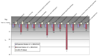
It is interesting that nebivolol but not atenolol leads to a downregulation of gene expression in glutamine-glutamate conversion and pyruvate
metabolism, the latter of particular interest in ischemia. Keywords: Atenolol,
flowchart,
gene expression,
Nebivolol
|
|
|
26. Gasser R;
Ablasser K;
Roessl U;
Kraigher-Krainer E;
Lewinski D;
Mangge H;
Mächler H;
Trantina-Yates A;
Tscheliessnigg KH;
Udermann H;
Porta S;
Friehs I;
Scherr E;
Brussee H;
Gasser S
Differential Gene Expression und Nebivolol and Atenolol during Experimental Ischemia in Human Myocardium
Well-oxygenized preparations
Journal of Clinical and Basic Cardiology 2008; 11 (1-4): 16-23
In well-oxygenized preparations, we can see that numerous biochemical processes are up-regulated by nebivolol (not by atenolol). Interesting are reverse transcription, stress response, and protein phosphorylation. Keywords: biochemical process,
flowchart
|
|
|
27. Gasser R;
Ablasser K;
Roessl U;
Kraigher-Krainer E;
Lewinski D;
Mangge H;
Mächler H;
Trantina-Yates A;
Tscheliessnigg KH;
Udermann H;
Porta S;
Friehs I;
Scherr E;
Brussee H;
Gasser S
Differential Gene Expression und Nebivolol and Atenolol during Experimental Ischemia in Human Myocardium
Biochemical process
Journal of Clinical and Basic Cardiology 2008; 11 (1-4): 16-23
In well-oxygenized preparations, we can see that numerous biochemical processes, mainly those involved in signalling and cellular immunity, are affected by nebivolol but not by atenolol. In the lower part, we can see that many processes involved in contraction, lipid metabolism, and proliferation are down-regulated by nebivolol only (not by atenolol). This kind of
processes, if slowed down, may render the heart less vulnerable to O2 deficit or ischemia. Keywords: biochemical process,
flowchart
|
|
|
28. Gasser R;
Ablasser K;
Roessl U;
Kraigher-Krainer E;
Lewinski D;
Mangge H;
Mächler H;
Trantina-Yates A;
Tscheliessnigg KH;
Udermann H;
Porta S;
Friehs I;
Scherr E;
Brussee H;
Gasser S
Differential Gene Expression und Nebivolol and Atenolol during Experimental Ischemia in Human Myocardium
Nebivolol - Atenolol
Journal of Clinical and Basic Cardiology 2008; 11 (1-4): 16-23
Similarly, as in Figure 4, biological processes involved in cell-mediated immunity and reverse transcription are significantly up-regulated by nebivolol and not by atenolol. One may deduce that both cellular immunity
as well as reverse transcription are regulated by nebivolol at various sites. Keywords: Atenolol,
flowchart,
Nebivolol
|
|
|
29. Gasser R;
Ablasser K;
Roessl U;
Kraigher-Krainer E;
Lewinski D;
Mangge H;
Mächler H;
Trantina-Yates A;
Tscheliessnigg KH;
Udermann H;
Porta S;
Friehs I;
Scherr E;
Brussee H;
Gasser S
Differential Gene Expression und Nebivolol and Atenolol during Experimental Ischemia in Human Myocardium
Nebivolol - Atenolol
Journal of Clinical and Basic Cardiology 2008; 11 (1-4): 16-23
This figure shows biological processes more than twofold downregulated in the presence of nebivolol but unaffected by atenolol during simulated ischemia (100 % O2 replaced by 100 % N2 perfusion). One can
see that, in particular on the level of reverse transcription and T-cell-mediated immunity as well as in the process of angiogenesis, a noteworthy regulation
is brought about by nebivolol. Keywords: Atenolol,
flowchart,
Nebivolol
|
|
|
30. Gasser R;
Ablasser K;
Roessl U;
Kraigher-Krainer E;
Lewinski D;
Mangge H;
Mächler H;
Trantina-Yates A;
Tscheliessnigg KH;
Udermann H;
Porta S;
Friehs I;
Scherr E;
Brussee H;
Gasser S
Differential Gene Expression und Nebivolol and Atenolol during Experimental Ischemia in Human Myocardium
Hypoxia
Journal of Clinical and Basic Cardiology 2008; 11 (1-4): 16-23
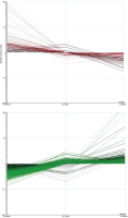
Panels A und B show all genes regulated significantly during experimental myocardial ischemia (hypoxia) in the presence and absence of either atenolol or nebivolol. Panel A (red) summarizes all
genes down-regulated by nebivolol compared to those up-regulated by atenolol during hypoxia. N2 control indicates gene expression in ischemic preparations without betablockers. N2 nebivolol indicates gene expression in the presence of nebivolol in ischemic preparations.
The red and green lines indicate the direction of gene regulation. On the left side of panel A, N2 atenolol represents genes upregulated under atenolol during hypoxia. Panel B, accordingly, shows all genes up-regulated by nebivolol which are down-regulated
by atenolol (N2 control here also representing non-treated controls). Keywords: Atenolol,
hypoxia,
Nebivolol
|
|
|
31. Gasser R;
Ablasser K;
Roessl U;
Kraigher-Krainer E;
Lewinski D;
Mangge H;
Mächler H;
Trantina-Yates A;
Tscheliessnigg KH;
Udermann H;
Porta S;
Friehs I;
Scherr E;
Brussee H;
Gasser S
Differential Gene Expression und Nebivolol and Atenolol during Experimental Ischemia in Human Myocardium
Atenolol - Nebivolol
Journal of Clinical and Basic Cardiology 2008; 11 (1-4): 16-23
Panels A und B show all genes regulated by either atenolol or nebivolol under non-ischemic, well-oxygenated conditions. Panel
A (red) summarizes all genes down-regulated by nebivolol compared to those up-regulated by atenolol during normoxia. Hence, panel A demonstrates the direct influence of atenolol and nebivolol
on gene expression in well-oxygenated preparations. It can easily be seen that, under optimal conditions, without any sign of ischemia/hypoxia, the effect of nebivolol upon gene expression is quite different
from that of atenolol. O2 control indicates gene expression in well-oxygenated preparations without betablockers. O2 nebivolol indicates gene expression in the presence of nebivolol in non-ischemic,
well-oxygenated preparations. The red and green lines indicate the direction of gene regulation. On the left side of panel A, O2 atenolol represents genes differentially expressed under atenolol during control
conditions. Panel B, accordingly, shows all genes up-regulated by nebivolol which are down-regulated by atenolol (O2 control here also representing untreated controls). Keywords: Atenolol,
Nebivolol
|
|
|
32. Gasser R;
Ablasser K;
Roessl U;
Kraigher-Krainer E;
Lewinski D;
Mangge H;
Mächler H;
Trantina-Yates A;
Tscheliessnigg KH;
Udermann H;
Porta S;
Friehs I;
Scherr E;
Brussee H;
Gasser S
Differential Gene Expression und Nebivolol and Atenolol during Experimental Ischemia in Human Myocardium
Micriarray experiments
Journal of Clinical and Basic Cardiology 2008; 11 (1-4): 16-23
Typical results derived from microarray experiments. Light intensities on the microtiter plate reflect gene expression. Keywords: light intensities
|
|
|
33. Gasser R;
Gasser S;
Ablasser K;
Scherr E;
Roessl U;
Lewinski D;
Mangge H;
Dellacher A;
Mächler H;
Trantina-Yates A;
Tscheliessnigg KH
Quantification of GLUT4 Gene Expression in Human Atrial Myocardium of Hypertensive Patients and the Effect of Experimental Ischaemia Thereupon
Expression of GLUT4
Journal of Clinical and Basic Cardiology 2007; 10 (1-4): 1-6
Relative difference in the expression of GLUT4 under ischaemic and non-ischaemic conditions (n = 8; ± SEM) in hypertensive, diabetic and control subjects. Results show slight trends in GLUT4 mRNA expression, however no statistical significance could be seen in either group. Keywords: Diagramm,
flowchart,
GLUT4,
ischaemia,
Ischämie
|
|
|
34. Gasser R;
Gasser S;
Ablasser K;
Scherr E;
Roessl U;
Lewinski D;
Mangge H;
Dellacher A;
Mächler H;
Trantina-Yates A;
Tscheliessnigg KH
Quantification of GLUT4 Gene Expression in Human Atrial Myocardium of Hypertensive Patients and the Effect of Experimental Ischaemia Thereupon
Hypertensive Patients
Journal of Clinical and Basic Cardiology 2007; 10 (1-4): 1-6
Mean ± SEM of the relative myocardial cellular GLUT4 gene expression in the three tested groups displays clearly the lower GLUT4 expression in hypertensive patients directly snap-frozen during cardiac surgery, not equilibrated with 100 % O2. Keywords: Diagramm,
flowchart,
GLUT4,
hypertension,
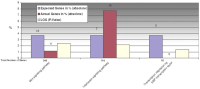
Hypertonie
|
|
|
35. Gasser R;
Gasser S;
Ablasser K;
Scherr E;
Roessl U;
Lewinski D;
Mangge H;
Dellacher A;
Mächler H;
Trantina-Yates A;
Tscheliessnigg KH
Quantification of GLUT4 Gene Expression in Human Atrial Myocardium of Hypertensive Patients and the Effect of Experimental Ischaemia Thereupon
GLUT4
Journal of Clinical and Basic Cardiology 2007; 10 (1-4): 1-6
The amount of cycles needed for the amplification curve to exceed the background fluorescence (dashed line) represents the CT value. The quantity of myocardial cellular GLUT4 gene expression is shown as relative difference, which is calculated from the CT values of the housekeeping and the GLUT4 PCR. Keywords: amplification curve,
Amplifikationskurve,
GLUT4
|
|
|
36. Castellani C;
Fruhwald FM;
Scherr D
NT-proBNP als Marker für eine erfolgreiche Therapie mit Levosimendan bei akuter Herzinsuffizienz. Fallbericht über einen Studenten aus dem Kosovo.
NT-proBNP
Journal für Kardiologie - Austrian Journal of Cardiology 2006; 13 (1-2): 37-39
Verlauf NT-proBNP. Rückgang des NT-proBNP von initial 30.888 pg/ml auf 7397 pg/ml nach Gabe von Levosimendan. Weitere Reduktion durch Medikation
und kardiale Resynchronisation (CRT) auf 462 pg/ml. Keywords: Diagramm,
Kardiologie,
Nt-proBNP-Spiegel
|
|
|
37. Castellani C;
Fruhwald FM;
Scherr D
NT-proBNP als Marker für eine erfolgreiche Therapie mit Levosimendan bei akuter Herzinsuffizienz. Fallbericht über einen Studenten aus dem Kosovo.
Leberparameter
Journal für Kardiologie - Austrian Journal of Cardiology 2006; 13 (1-2): 37-39
Verlauf der Leberparameter in den ersten Tagen: deutlicher Rückgang der Transaminasen und der LDH innerhalb der ersten 10 Tage. Keywords: Diagramm,
Enzymspiegel,
Kardiologie
|
|
|
38. Scherr D
Überlebter plötzlicher Herzkreislaufstillstand bei einem Patienten ohne strukturelle Herzerkrankung: das Brugada-Syndrom
Brugada-Syndrom - Ajmalin
Journal für Kardiologie - Austrian Journal of Cardiology 2003; 10 (Supplementum B - Forum Rhythmologie): 8-10
Das 12-Kanal-EKG nach Gabe von 1 mg/kg Ajmalin: Entwicklung der für das Brugada-Syndrom typischen J-Punkt-Hebung und deszendierenden ST-Streckenhebung mit nachfolgender negativer T-Welle in den rechtspräkordialen Ableitungen. Keywords: Ajmalin,
Brugada-Syndrom,
EKG,
Elektrokardiographie,
Fallbericht,
J-Punkt-Hebung,
Kardiologie,
ST-Streckenhebung
|
|
|
39. Scherr D
Überlebter plötzlicher Herzkreislaufstillstand bei einem Patienten ohne strukturelle Herzerkrankung: das Brugada-Syndrom
Brugada-Syndrom - Ajmalin-Test
Journal für Kardiologie - Austrian Journal of Cardiology 2003; 10 (Supplementum B - Forum Rhythmologie): 8-10
Das 12-Kanal-EKG vor der Durchführung des Ajmalin-Tests. Keywords: Ajmalin-Test,
Brugada-Syndrom,
EKG,
Elektrokardiographie,
Fallbericht,
Kardiologie
|
|
|
40. Scherr D
Überlebter plötzlicher Herzkreislaufstillstand bei einem Patienten ohne strukturelle Herzerkrankung: das Brugada-Syndrom
Brugada-Syndrom - EKG
Journal für Kardiologie - Austrian Journal of Cardiology 2003; 10 (Supplementum B - Forum Rhythmologie): 8-10
Das 12-Kanal-EKG des Patienten bei Aufnahme zeigt einen rechtsschenkelblockartig deformierten Kammerkomplex (J-Punkt-Hebung in V1 und V2) und eine deszendierende ST-Streckenhebung in den Ableitungen V1 und V2. Keywords: Brugada-Syndrom,
EKG,
Elektrokardiographie,
Fallbericht,
J-Punkt-Hebung,
Kammerkomplex,
Kardiologie,
Streckenhebung
|
|
|
| |

 Suche Medizinische Publikationen
 Suche Abbildungen und Grafiken
 Medizinprodukte - Labortechnik
 Antidotarium (Stand 2012)
 Phytokodex (Stand 2012)
 Fachbücher
 Patientenliteratur