|
|
|---|
3561. Abacioglu U
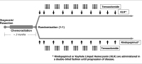
Interview with Dr Michael Weller (Zurich) about the Phase-III EGFRvIII Vaccine Trial on Newly Diagnosed EGFRvIII-Positive Glioblastoma
ACT IV
European Association of NeuroOncology Magazine 2013; 3 (3): 155-156
Trial scheme Keywords: ACT IV,
scheme
|
|
|
3562. Florian IA
Congress Report: 3rd National Conference of Neuro-Oncology with International Participation
Congress Report
European Association of NeuroOncology Magazine 2013; 3 (3): 150
Closing ceremony. |
|
|
3563. Zijdewind J;
Wesseling P;
de Witt Hamer PC;
Reijneveld J
Case Report: Primary Leptomeningeal Melanoma in a Patient with Neurocutaneous Melanosis
Leptomeningeal Tumour
European Association of NeuroOncology Magazine 2013; 3 (3): 141-142
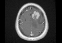
Transversal section of contrast-enhanced T1 MRI demonstrating a left frontal, inhomogeneously contrast-enhancing, and space-occupying lesion with oedema in the surrounding brain tissue. Keywords: leptomeningeal tumour,
MRI
|
|
|
3564. Zijdewind J;
Wesseling P;
de Witt Hamer PC;
Reijneveld J
Case Report: Primary Leptomeningeal Melanoma in a Patient with Neurocutaneous Melanosis
Leptomeningeal Tumour
European Association of NeuroOncology Magazine 2013; 3 (3): 141-142
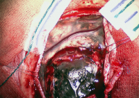
Intraoperative photograph after opening of the dura, showing a black leptomeningeal tumour in the left frontal region. Of note is the spotty black
appearance of the arachnoid, which is not continuous with the tumour, consistent with neurocutaneous melanosis. Keywords: leptomeningeal tumour,
photo
|
|
|
3565. Rössler K;
Coras R;
Blümcke I
Case Report:Long-Term Epilepsy-Associated Giant Lobar Collision Tumour
Brain-Collision-Tumour
European Association of NeuroOncology Magazine 2013; 3 (3): 139-140
Ependymoma component (H & E staining). Keywords: ependymoma,
histology
|
|
|
3566. Rössler K;
Coras R;
Blümcke I
Case Report:Long-Term Epilepsy-Associated Giant Lobar Collision Tumour
Brain-Collision-Tumour
European Association of NeuroOncology Magazine 2013; 3 (3): 139-140
Higher magnification of ganglioglioma component with dysplastic neurons (H & E staining). Keywords: ganglioglioma,
histology
|
|
|
3567. Rössler K;
Coras R;
Blümcke I
Case Report:Long-Term Epilepsy-Associated Giant Lobar Collision Tumour
Brain-Collision-Tumour
European Association of NeuroOncology Magazine 2013; 3 (3): 139-140
Ganglioma component (H & E staining). Keywords: ganglioglioma,
histology
|
|
|
3568. Rössler K;
Coras R;
Blümcke I
Case Report:Long-Term Epilepsy-Associated Giant Lobar Collision Tumour
Brain-Collision-Tumour
European Association of NeuroOncology Magazine 2013; 3 (3): 139-140
Increased proliferation activity as indexed by MIB1 labelling. Keywords: histology,
neuroepithelial tumour
|
|
|
3569. Rössler K;
Coras R;
Blümcke I
Case Report:Long-Term Epilepsy-Associated Giant Lobar Collision Tumour
Brain-Collision-Tumour
European Association of NeuroOncology Magazine 2013; 3 (3): 139-140
Macroscopic appearance of the tumour. Keywords: photo,
tumour
|
|
|
3570. Grisold W;
Soffietti R
Editorial to Special Issue
Nerve-Infiltration
European Association of NeuroOncology Magazine 2013; 3 (3): 94
Spinal nerve root in a patient with lung cancer. Tumour tissue within the nerve root (arrow). Keywords: malignant tumour,
nerve infiltration
|
|
|
3571. Grisold W;
Soffietti R
Editorial to Special Issue
Nerve-Infiltration
European Association of NeuroOncology Magazine 2013; 3 (3): 94
Infiltration of a trigeminal branch in the cavernous sinus by a gliosarcoma (arrow). Keywords: malignant tumour,
nerve infiltration
|
|
|
|
|
|
3573. Rössler K;
Coras R;
Blümcke I
Case Report:Long-Term Epilepsy-Associated Giant Lobar Collision Tumour
Brain-Collision-Tumour
European Association of NeuroOncology Magazine 2013; 3 (3): 139-140
T2 axial MRI, calcification in black. Keywords: intrinsic tumour,
left frontal lobe,
MRI
|
|
|
3574. Rössler K;
Coras R;
Blümcke I
Case Report:Long-Term Epilepsy-Associated Giant Lobar Collision Tumour
Brain-Collision-Tumour
European Association of NeuroOncology Magazine 2013; 3 (3): 139-140
T1 gadolinium-enhanced axial MRI. Keywords: intrinsic tumour,
left frontal lobe,
MRI
|
|
|
3575. Rössler K;
Coras R;
Blümcke I
Case Report:Long-Term Epilepsy-Associated Giant Lobar Collision Tumour
Brain-Collision-Tumour
European Association of NeuroOncology Magazine 2013; 3 (3): 139-140
Cranial CT scan at the age of 14 months. Keywords: CT,
porencephalic cyst
|
|
|
3576. Perneczky G;
Loyoddin M;
Schappelwein H;
Sherif C
Spinal Meningiomas: A Comprehensive Overview and Own Experience
Spinal Menigioma
European Association of NeuroOncology Magazine 2013; 3 (3): 118-121
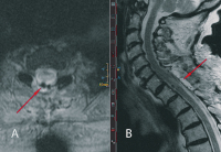
Postoperative MR scans: (a) T1-weighted contrast-enhanced MR images: the spinal cord (red arrow) is in normal location. No tumour remnants are visible. (b) T2-weighted native MR scan: the surgical dorsal approach and complete tumour
removal are documented. Keywords: MRI,
spinal meningioma
|
|
|
3577. Perneczky G;
Loyoddin M;
Schappelwein H;
Sherif C
Spinal Meningiomas: A Comprehensive Overview and Own Experience
Spinal Meningioma
European Association of NeuroOncology Magazine 2013; 3 (3): 118-121
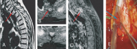
Pre- and intraoperative findings: (a) T2-weighted native sagittal MR image: the tumour can be nicely delineated (red arrow). (b, c) Axial T1-weighted contrastenhanced sequence: inhomogeneous contrast enhancement is due to intratumoural calcifications (red arrow). (d) Sagittal contrast-enhanced T1-weighted image: again, the calcifications can be seen as contrast agent-free areas within the tumour (red arrow). (e) Intraoperative findings: the spinal cord is carefully mobilized under IOM. The tumour (blue arrow) can be seen at the left ventral dura. Keywords: MRI,
spinal meningioma
|
|
|
3578. Millesi M;
Kueß M;
Widhalm G;
Knosp E
Current Surgical Strategies in the Treatment of Intracranial Meningiomas
Intracranial Meningioma
European Association of NeuroOncology Magazine 2013; 3 (3): 112-117
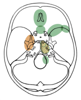
Current treatment strategies for skull base meningiomas. Green: surgical resection; brown:
radiosurgery; green-brown: the best therapeutic option for petroclival meningiomas is still a matter of discussion. Keywords: scheme,
skull base meningioma,
treatment strategy
|
|
|
3579. Millesi M;
Kueß M;
Widhalm G;
Knosp E
Current Surgical Strategies in the Treatment of Intracranial Meningiomas
Intracranial Meningioma
European Association of NeuroOncology Magazine 2013; 3 (3): 112-117
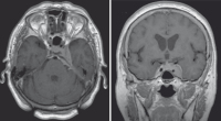
Left-sided cavernous-sinus meningioma found due to double vision treated by radiosurgery. Keywords: cavernous-sinus meningioma
|
|
|
3580. Millesi M;
Kueß M;
Widhalm G;
Knosp E
Current Surgical Strategies in the Treatment of Intracranial Meningiomas
Intracranial Meningioma
European Association of NeuroOncology Magazine 2013; 3 (3): 112-117
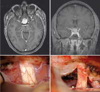
Tuberculum sellae meningioma found due to blurred
vision operated via a right-sided subfrontal approach. Keywords: tuberculum sellae meningioma
|
|
|
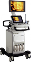
3581. Medizinprodukte - Labortechnik: Der neue UGEO H60 - Einzigartige Bildqualität und maximale Diagnosesicherheit
Produkt: UGEO H60
Vertrieb: Mides GmbH
Journal für Gynäkologische Endokrinologie 2013; 7 (3) (Ausgabe für Österreich): 26
Keywords: Gynäkologie-MT,
Mides GmbH,
Reproduktion,
Reproduktionsmedizin,
Reproduktionsmedizin-MT,
UGEO H60
|
|
|
3582. Brezinka C
Tipps und Tricks im Gyn-Ultraschall: Das prämenstruelle und das menstruelle Endometrium
Gyn-Ultraschall
Journal für Gynäkologische Endokrinologie 2013; 7 (3) (Ausgabe für Österreich): 24-25
Journal für Gynäkologische Endokrinologie 2013; 7 (3) (Ausgabe für Schweiz): 33-34
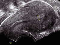
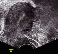
Prämenstruelles Bild in der 2. Zyklushälfte: Stratum basale und Stratum functionale sind nicht mehr zu unterscheiden, auch die mediane Linie ist nicht mehr sichtbar. Die abgegriffene Strecke ist 11 mm breit (D-Muster nach Lindenberg). Keywords: Endometrium,
Gynäkologie,
Menstruation,
Ultraschall,
Zyklus
|
|
|
3583. Brezinka C
Tipps und Tricks im Gyn-Ultraschall: Das prämenstruelle und das menstruelle Endometrium
Gyn-Ultraschall
Journal für Gynäkologische Endokrinologie 2013; 7 (3) (Ausgabe für Österreich): 24-25
Journal für Gynäkologische Endokrinologie 2013; 7 (3) (Ausgabe für Schweiz): 33-34
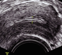
Ab dem 5. Zyklustag entsteht aus dem bisher diffusen Bild eine Struktur mit einem markanten Mittelecho (B-Muster nach Lindenberg). Die abgegriffene Strecke ist 3 mm breit. Keywords: Endometrium,
Gynäkologie,
Menstruation,
Ultraschall,
Zyklus
|
|
|
3584. Brezinka C
Tipps und Tricks im Gyn-Ultraschall: Das prämenstruelle und das menstruelle Endometrium
Gyn-Ultraschall
Journal für Gynäkologische Endokrinologie 2013; 7 (3) (Ausgabe für Österreich): 24-25
Journal für Gynäkologische Endokrinologie 2013; 7 (3) (Ausgabe für Schweiz): 33-34
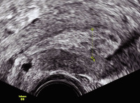
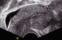
Das menstruelle Endometrium muss nicht immer ein dünner Strich sein, eine eher diffuse Zone von 4 mm Breite wie in diesem Bild ist ein häufiger
Befund (A-Muster nach Lindenberg). Keywords: Endometrium,
Gynäkologie,
Menstruation,
Ultraschall,
Zyklus
|
|
|
3585. Brezinka C
Tipps und Tricks im Gyn-Ultraschall: Das prämenstruelle und das menstruelle Endometrium
Gyn-Ultraschall
Journal für Gynäkologische Endokrinologie 2013; 7 (3) (Ausgabe für Österreich): 24-25
Journal für Gynäkologische Endokrinologie 2013; 7 (3) (Ausgabe für Schweiz): 33-34
Dünnes, diffuses Endometrium mit einer Breite von 2 mm ist der häufigste Befund in den ersten beiden Tagen des Zyklus (A-Muster nach Lindenberg). Keywords: Endometrium,
Gynäkologie,
Menstruation,
Ultraschall,
Zyklus
|
|
|
3586. Brezinka C
Tipps und Tricks im Gyn-Ultraschall: Das prämenstruelle und das menstruelle Endometrium
Gyn-Ultraschall
Journal für Gynäkologische Endokrinologie 2013; 7 (3) (Ausgabe für Österreich): 24-25
Journal für Gynäkologische Endokrinologie 2013; 7 (3) (Ausgabe für Schweiz): 33-34
Ein kaum sichtbarer, nicht genau abgrenzbarer Streifen (A-Muster nach Lindenberg) lässt das Cavum uteri nur erahnen; gleichzeitig im Douglas ein 6 mm breiter Flüssigkeitssaum bei retrograder Menstruation. Keywords: Endometrium,
Gynäkologie,
Menstruation,
Ultraschall,
Zyklus
|
|
|
3587. Brezinka C
Tipps und Tricks im Gyn-Ultraschall: Das prämenstruelle und das menstruelle Endometrium
Gyn-Ultraschall
Journal für Gynäkologische Endokrinologie 2013; 7 (3) (Ausgabe für Österreich): 24-25
Journal für Gynäkologische Endokrinologie 2013; 7 (3) (Ausgabe für Schweiz): 33-34
Am 2. Zyklustag kann sich manchmal ein echoarmer Streifen im Sinne einer Serometra zeigen (M-Muster nach Lindenberg). Die abgegriffene Strecke ist 3 mm breit. Keywords: Endometrium,
Gynäkologie,
Menstruation,
Ultraschall,
Zyklus
|
|
|
3588. Brezinka C
Tipps und Tricks im Gyn-Ultraschall: Das prämenstruelle und das menstruelle Endometrium
Gyn-Ultraschall
Journal für Gynäkologische Endokrinologie 2013; 7 (3) (Ausgabe für Österreich): 24-25
Journal für Gynäkologische Endokrinologie 2013; 7 (3) (Ausgabe für Schweiz): 33-34
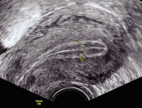
Um die Ovulation zeigt sich eine deutliche ā€žtriple lineā€œ. Das Stratum basale ist echodens und hebt sich sichtlich gegenĆ¼ber dem Endometrium ab, die zentrale 3. Linie wird durch das Aufeinanderliegen der beiden Schichten des Stratum functionale
gebildet (C-Muster nach Lindenberg). Die abgegriffene Strecke ist 10 mm breit. Keywords: Endometrium,
Gynäkologie,
Ovulation,
Ultraschall,
Zyklus
|
|
|
3589. Prückler J;
Leitner G;
Klein M;
Gruber I
Ernährungstherapie und Essverhalten bei Polyzystischem Ovarialsyndrom (PCOS) im Rahmen einer Kinderwunschbehandlung
Essverhalten-PCOS
Journal für Gynäkologische Endokrinologie 2013; 7 (3) (Ausgabe für Österreich): 14-17
Journal für Gynäkologische Endokrinologie 2013; 7 (3) (Ausgabe für Schweiz): 11-14
Body-Mass-Index (Median) von Frauen mit PCOS (n = 11) im Verlauf. Keywords: BMI,
Body-mass-Index,
Diagramm,
Gewichtsverlauf,
Gynäkologie,
PCOS
|
|
|
3590. Köhn FM;
Schuppe H-C;
Haidl G
Unerfüllter Kinderwunsch: Welche Rolle spielt der Mann?
SCO-Syndrom
Journal für Urologie und Urogynäkologie 2013; 20 (3) (Ausgabe für Österreich): 12-21
Journal für Urologie und Urogynäkologie 2013; 20 (3) (Ausgabe für Schweiz): 10-19
Testikuläres Gewebe mit (a) ausreifender Spermato- und Spermiogenese mit reifen Spermatiden und (b) Sertoli-cell-only-Syndrom ohne Nachweis eines Keimepithels. Keywords: Andrologie,
Germinalzellaplasie,
SCO-Syndrom,
Spermatogenese
|
|
|
3591. Kufner M;
Kruck S;
Nagele U
Vergleich zwischen Stoßwellenlithotripsie (ESWL), Ureterorenoskopie (URS) und minimalinvasiver perkutaner Nephrolitholapaxie (PCNL/MIP) zur Behandlung der Nephrolithiasis
Nephrolithiasis
Journal für Urologie und Urogynäkologie 2013; 20 (3) (Ausgabe für Österreich): 5-9
Journal für Urologie und Urogynäkologie 2013; 20 (3) (Ausgabe für Schweiz): 4-7
Vergleich Steinrezidivrate im zeitlichen Verlauf. UKG: untere Kelchgruppe; ESWL: Stoßwellenlithotripsie; MIP: minimalinvasive perkutane Nephrolitholapaxie; URS: Ureterorenoskopie. Keywords: Diagramm,
ESWL,
MIP,
Steinrezidivrate,
UKG,
URS,
zeitlicher Verlauf
|
|
|
3592. Kufner M;
Kruck S;
Nagele U
Vergleich zwischen Stoßwellenlithotripsie (ESWL), Ureterorenoskopie (URS) und minimalinvasiver perkutaner Nephrolitholapaxie (PCNL/MIP) zur Behandlung der Nephrolithiasis
Nephrolithiasis
Journal für Urologie und Urogynäkologie 2013; 20 (3) (Ausgabe für Österreich): 5-9
Journal für Urologie und Urogynäkologie 2013; 20 (3) (Ausgabe für Schweiz): 4-7
Vergleich Steingröße und Steinfreiheitsrate zwischen den unterschiedlichen Behandlungsmodalitäten. UKG: untere Kelchgruppe; ESWL: Stoßwellenlithotripsie; MIP: minimalinvasive perkutane Nephrolitholapaxie; URS: Ureterorenoskopie. Keywords: Diagramm,
ESWL,
MIP,
Steinfreiheitsrate,
Steingröße,
UKG,
URS
|
|
|
3593. Schmid M
Klinische Anwendung von Microarrays (ā€žGenchipsā€œ) in der PrƤnataldiagnostik
Microarrays
Speculum - Zeitschrift für Gynäkologie und Geburtshilfe 2013; 31 (3) (Ausgabe für Österreich): 18-21
Speculum - Zeitschrift für Gynäkologie und Geburtshilfe 2013; 31 (3) (Ausgabe für Schweiz): 20-23
Empfehlung zur klinischen Anwendung der Chromosomen-Microarray-Analyse in der Pränataldiagnostik. CVS: Chorionzottenbiopsie; AC: Amniozentese; NT: Nackentransparenz. Keywords: Chromosomen-Microarray-Analyse,
CMA,
Schema,
Speculum
|
|
|
|
|
|
|
|
|
|
|
|
|
|
|
|
|
|
3599. Aichholzer M;
Tomancok B;
Wies W;
Wurm G;
Sonnberger M
Ein Jahr Erfahrung mit dem intraoperativen Hochfeld-MRT
iMRT
Journal für Neurologie, Neurochirurgie und Psychiatrie 2013; 14 (3): 114-118
45-jähriger Patient mit epileptischem Anfall 2009, offener Biopsie am Tumor im Inselbereich 2009 (fibrilläres Astrozytom WHO II), Häufung der epileptischen Anfälle und Tumorwachstum in den MRT-Kontrollen; (a) präoperativ 09/2011 (2× axial
Flair/T1 + Kontrastmittel [KM]) mit neuer KM-Aufnahme links frontal; (b) intraoperativ 09/2011 (axial Flair/T1 + KM): KM-affiner Herd ist reseziert, aber noch ausgedehnter Tumorrest in den Randbereichen; (c) 3 Monate postoperativ 03/2012 (2× axial Flair/T1 + KM): geringe Resttumoranteile in den temporodorsalen Anteilen (Histologie:
anaplastisches Astrozytom WHO III). Keywords: Astrozytom III,
Hirntumor,
iMRT,
MRT
|
|
|
3600. Aichholzer M;
Tomancok B;
Wies W;
Wurm G;
Sonnberger M
Ein Jahr Erfahrung mit dem intraoperativen Hochfeld-MRT
iMRT
Journal für Neurologie, Neurochirurgie und Psychiatrie 2013; 14 (3): 114-118
(a) Hypophysenadenom präoperativ 11/2011 (T1 + Kontrastmittel [KM]): zystisch solides Hypophysenadenom mit parasellärer Tumorausdehnung rechts; (b) intraoperativ 11/2011 (T1 + KM): mit kleinem residualem Tumorausläufer an der medialen Sinus-cavernosus-Wand rechts; (c) postoperativ 02/2012 (T1 + KM): Bestätigung der kompletten Tumorresektion in der postoperativen 3-Monats-Kontrolle. Keywords: Hypophysenadenom,
Hypophysenchirurgie,
iMRT,
MRT
|
|
|
| |

 Suche Medizinische Publikationen
 Suche Abbildungen und Grafiken
 Medizinprodukte - Labortechnik
 Antidotarium (Stand 2012)
 Phytokodex (Stand 2012)
 Fachbücher
 Patientenliteratur